7 Feb 2023
Kate Parkinson BVSc, MRCVS shares a case study on the treatment of severe bilateral forelimb trauma, including management and discussion.
Kate Parkinson
Job Title
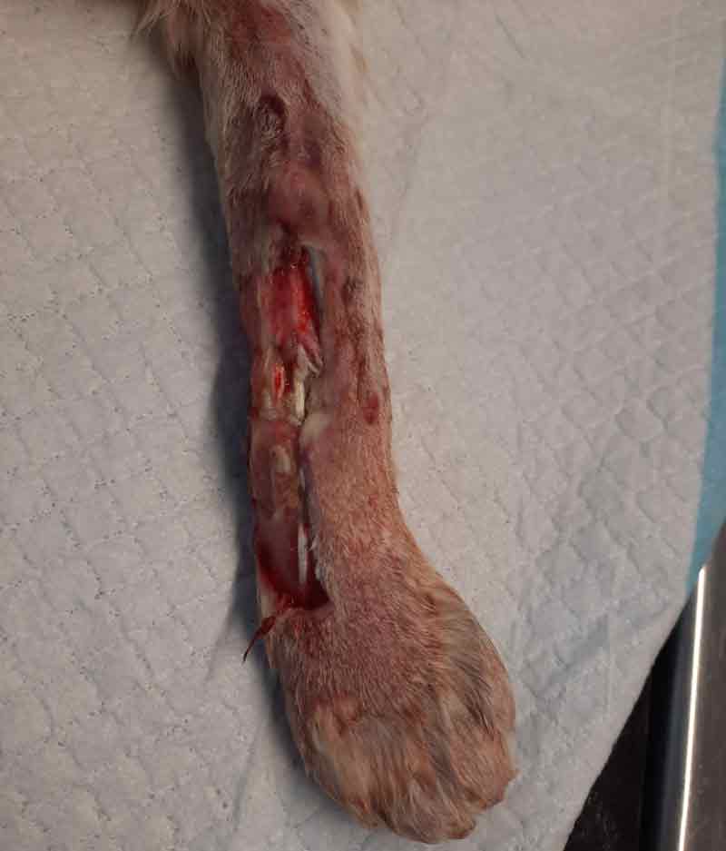
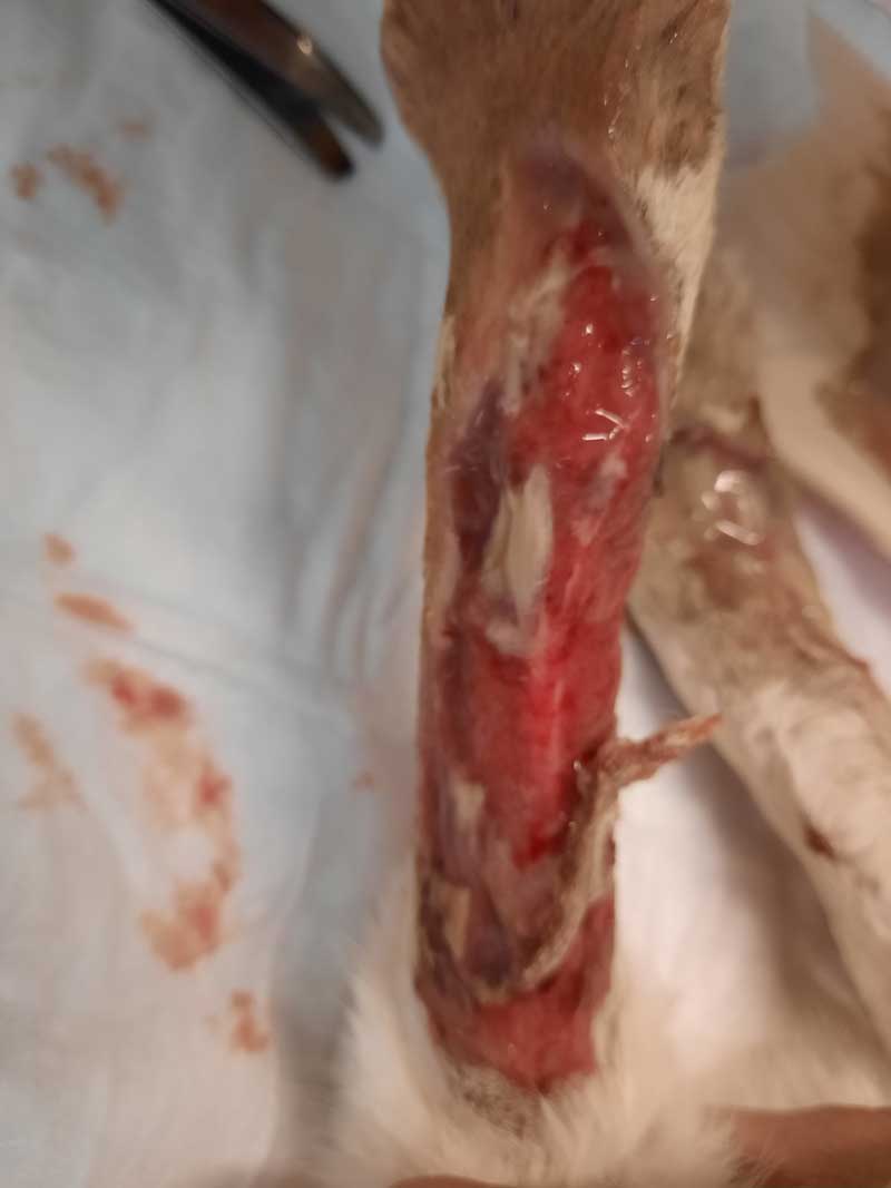
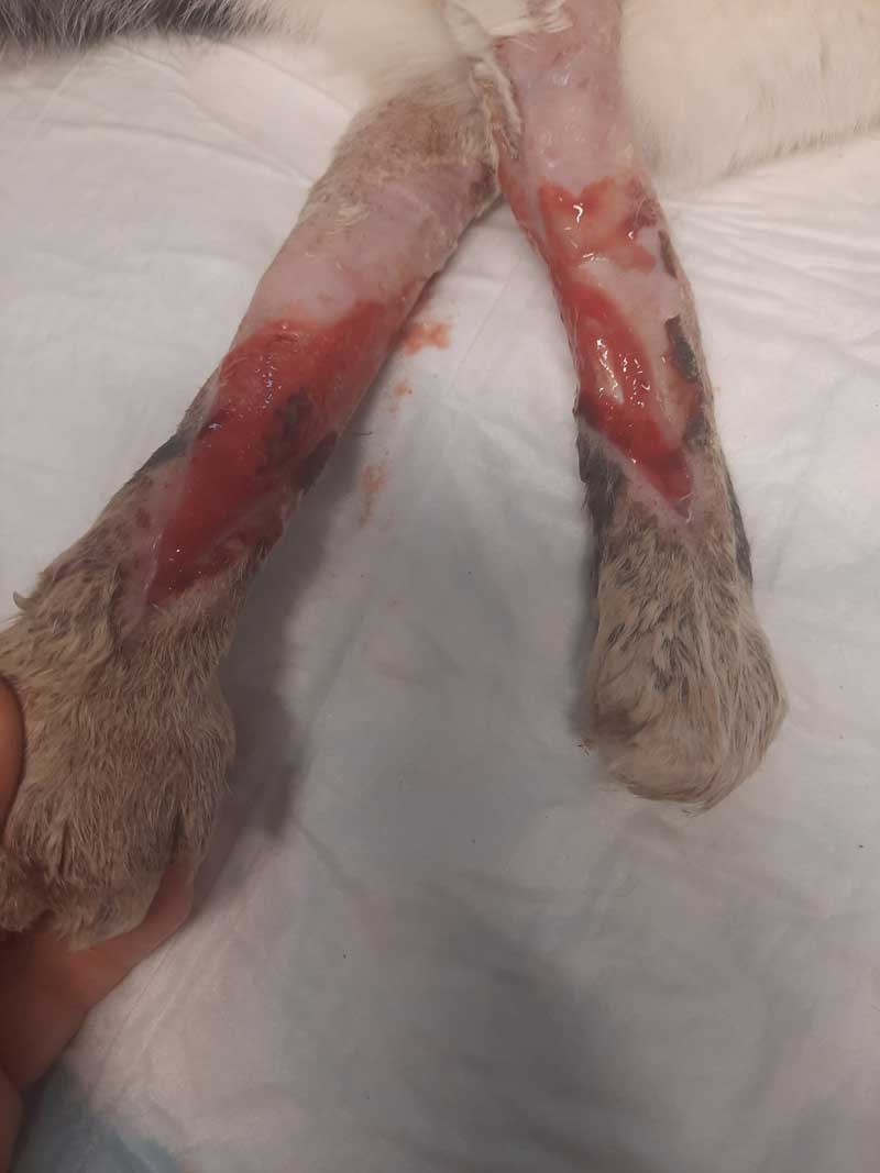
Figure 1. Left forelimb on presentation.
Wounds are a common presentation in first opinion feline small animal practice. This case study focuses on a severe bilateral forelimb degloving injury in a 10- year-old domestic shorthaired cat, which healed completely within two months using a variety of wound management techniques.
As wound healing occurs in three overlapping phases – the immediate, inflammatory phase; the proliferative or repair phase; and the remodelling phase – different approaches were required for the different phases. During the inflammatory phase, treatment was focused on wound debridement, which in this case was achieved by mechanical (wet to dry dressings, wound lavage) and surgical means (debridement). In the proliferative phase, treatment is focused on the formation and retention of healthy granulation tissue. Alginate dressings were used in this case to keep the wound environment moist and undisturbed. During remodelling, silver sulfadiazine cream was utilised to prevent infection and reduce over-granulation.
Although at times challenging, this case was well within the resources of first opinion practice, and the wounds healed well due to the cat’s exceptional temperament and its owner’s commitment.
A 10-year-old female neutered domestic shorthaired cat was presented as an emergency to the practice.
Milly was reported to have gone out that morning, then presented collapsed four hours later at the owners’ back door. On initial triage, the cat was in lateral recumbency with pale pink mucous membranes, a heart rate of 210bpm and a respiratory rate of 60. Her temperature was 38.3°C, but her extremities were cold.
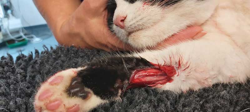
She had severe degloving injuries to both front legs (Figures 1 and 2), and a small wound over her left hock. Her injuries were thought likely to be consistent with being hit by a car.
Analgesia was given immediately (methadone 0.3mg/kg IM). Warming measures – blankets, heat pad and temperature management unit – were implemented. The injuries to the cat’s limbs made initial IV access challenging, but an IV catheter was inserted into her left femoral vein. A bolus of 10ml/kg warmed isotonic saline was administered over 10 minutes.
The patient’s injuries were severe, though not immediately life threatening, and potentially consistent with a guarded prognosis or lengthy recovery. This was discussed with the cat’s owners and permission was gained for initial investigations.
Conscious survey radiographs were taken, revealing no visible fractures. Shallow corneal ulcers were noted in both eyes, possibly due to trauma, and lubrication was applied.
Once the patient was stable, she was sedated to clip and clean her injuries. Both forelegs were extensively degloved. The foreleg skin was extremely bruised and was thought likely to devitalise. Due to this, the initial plan of attempting a surgical repair was abandoned. Both hindlegs also had minor injuries.
The skin of all four feet was clipped and the wounds flushed with saline. Sterile gauze swabs were moistened with sterile isotonic saline and applied directly to the wound as wet to dry dressings. A three-layer closure of padded, conforming and cohesive bandage was applied.
Anti-inflammatories (meloxicam 0.1mg/kg) and broad-spectrum antibiotic cover (amoxicillin-clavulanic acid, 8.75mg/kg) were administered by SC injection. Ofloxacin eye drops (0.3%) were started in both eyes four times daily and opioid pain relief was continued, with methadone injections every four hours. IV fluids were continued.
The owners were updated at this point and the cat was hospitalised. Referral was discussed at this point, but declined.
The following day, the cat was non-ambulatory, but was rolling over in her cage. She had eaten and was bright. She had not urinated, but her bladder was small and soft. She was sedated with a medetomidine/butorphanol combination.
The wounds were debrided and flushed with sterile saline, and the wet-to-dry dressings were replaced. Her bladder was expressed under sedation. The author’s team decided to continue with medication and reassess the wounds in 24 hours.
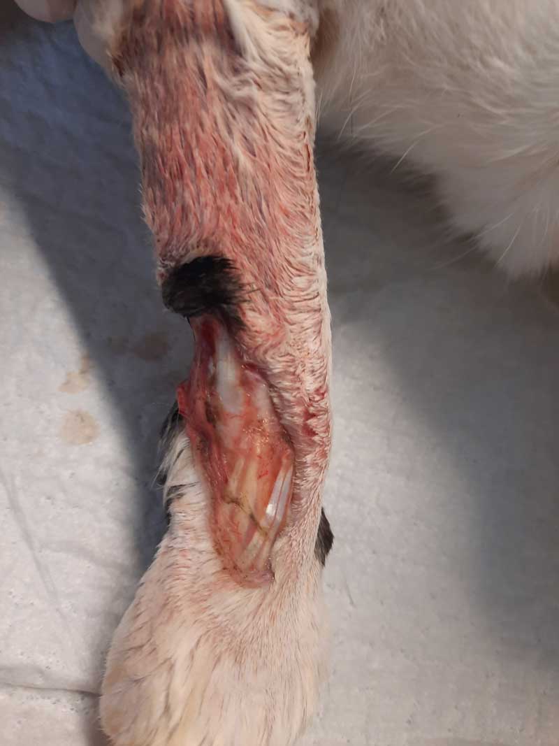
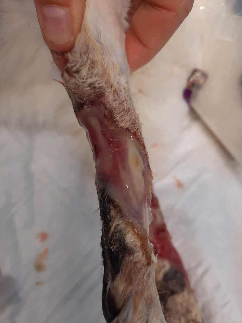
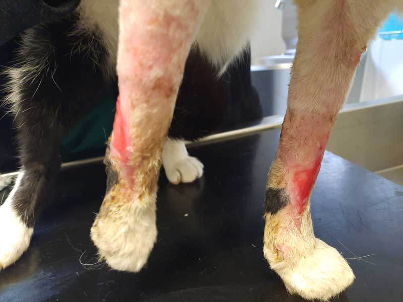
The next day, the cat was again bright, but non-ambulatory, and had not urinated. Necrotic tissue was removed from both forelimbs under sedation, leaving significant skin deficits and tendons exposed. (Figures 3 and 4). The hindfeet pads were warm and pink, but the front pads paler and cold. The wounds were redressed with wet-to-dry dressings and her bladder was expressed. The owners visited to discuss the case, and photographs of the wounds were shown. The decision was made to continue with treatment.
Sedated redressing was continued. The wounds were debrided and showed a small improvement, with healthy granulation tissue beginning to form. The bladder was expressed.
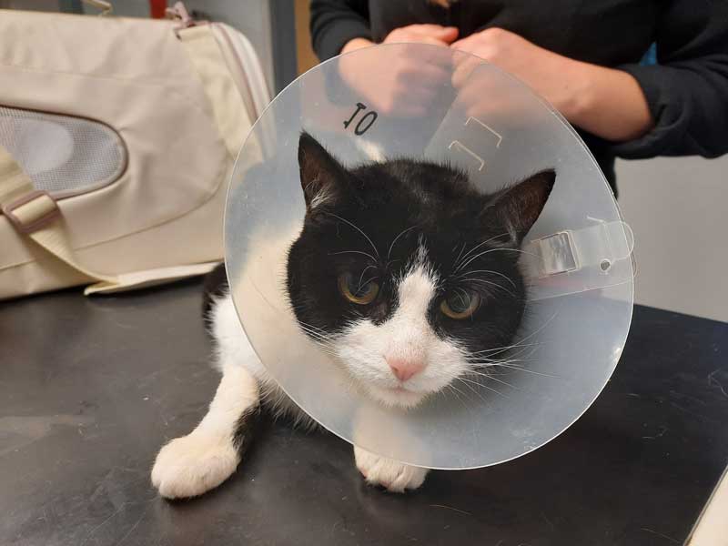
The decision was made to discharge the cat for 24 hours to see how she coped at home and to repeat the dressing in two days. The cat was discharged with oral meloxicam (0.2mg/kg by mouth once a day), transmucosal buprenorphine (0.02mg/kg three times a day), oral amoxicillin-clavulanate (12.5mg/kg twice a day), and intraocular ofloxacin (one drop four times a day). An Elizabethan collar was added as the cat was now more mobile.
Sedated redressing was repeated. Due to the large amount of necrotic tissue on the bandages, medical grade manuka honey and non-adhesive absorbent foam dressings were used beneath a three-layer bandage dressing. By now, the small wound on the left hindleg had healed. The cat was eating well at home, but was non-ambulatory.
She was also pyrexic (39.4°C) and was not urinating voluntarily, which could have been due to nerve damage or her lack of ambulation.
The cat’s dressings were changed in 24 hours to see how wound healing was progressing. A significant amount of necrotic tissue was removed.
The left eye ulcer had healed completely and the ulcer in her right eye was much smaller, so the ofloxacin drops were continued in the right eye only. Her bladder was again expressed, and all medical treatment was continued. Redressing was planned in 48 hours.
The cat re-presented 24 hours later. She had urinated at night and was much more ambulatory at home. However, her Elizabethan collar had been removed and she had chewed her dressings. Both foreleg dressings had slipped.
The forelimb dressings were removed, the wounds flushed with sterile saline, and the dressings replaced conscious.
The cat was sedated again for debridement and dressing change. Although peripherally, the wounds were granulating well, the removal of more necrotic tissue over the right carpus left bone exposed and the tendons were starting to fray.
The wounds were debrided, copiously lavaged and redressed with manuka honey and non-adhesive foam dressings as before. It was decided to redress again in 48 hours as the owners were concerned with the potential effect of repeated sedations.
On arrival for redressing, the owners reported the cat had not been walking as much and had not urinated that day. The dressings were removed conscious and the cat was placed on the ground to see if she could walk, which she did.
She was sedated and the dressings removed. The left forefoot was swollen and discoloured with a significant amount of necrotic skin around the edges of the wound.
The right front paw was not swollen, but a significant amount of necrotic tissue and discharge was present. Concerns were discussed with the owner and photographs were shown.
Daily sedation was reinstituted, and honey and non-adhesive foam dressings were applied beneath a three-layer bandage.
Oral amoxicillin-clavulanate was replaced with a second-line antibiotic (marbofloxacin 2mg/kg by mouth once a day).
The eye ulcers had healed, so ofloxacin was discontinued. The bladder was expressed.
The owners reported that the cat had improved and had passed faeces voluntarily, but not urine.
She was walking on arrival, and was bright and responsive. Sedated debridement and redressing was repeated.
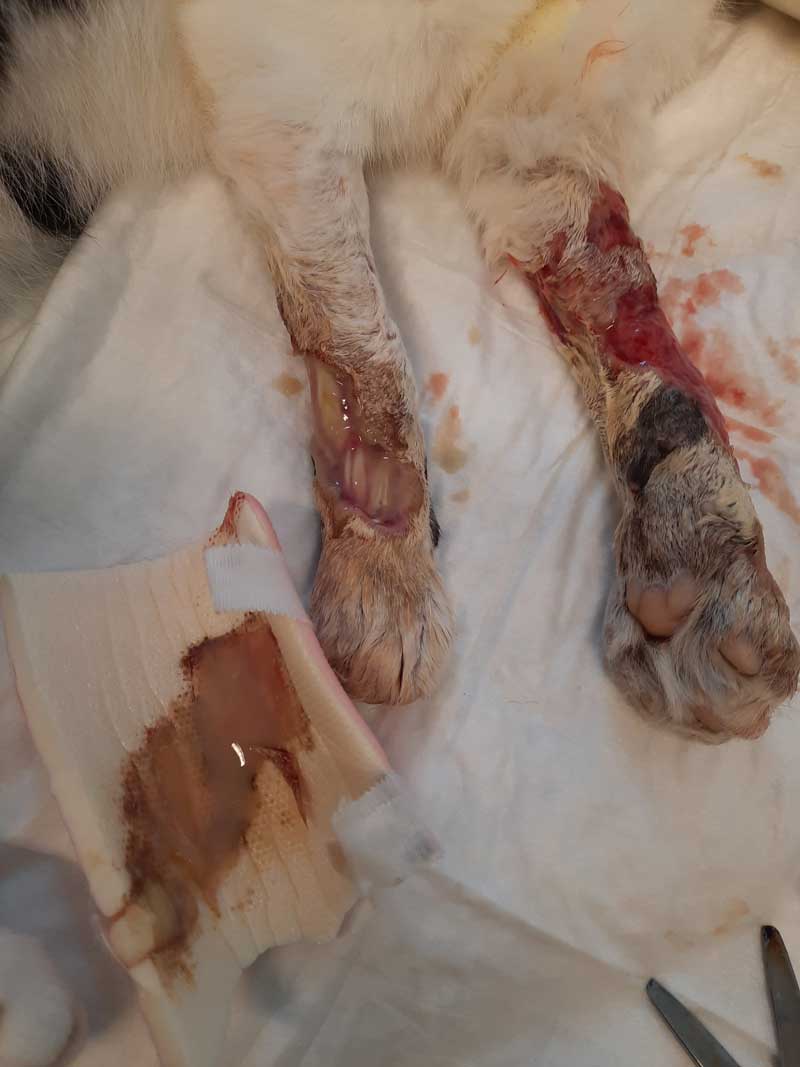
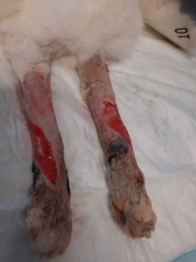
The swelling noted the previous day on the left foreleg had improved, and less discharge and necrotic tissue was present on the dressings (Figures 5 to 7).
The cat was still constipated and an enema was also performed. The bladder was again expressed. Oral buprenorphine was reduced to twice daily in the hope this would aid the passage of faeces.
Daily dressings under sedation with honey and non-adhesive foam dressing beneath a three-layer bandage were continued on days 12, 13 and 14.
By day 12, the cat was using her litter tray, and had passed both faeces and urine.
She was still slightly lame on her left leg compared to the right, but was walking.
On days 13 and 14, more necrotic tissue was removed. The wounds were still deep and an increasing amount of exudate was present on the dressings.
Referral was again declined, but permission was granted to discuss the case with colleagues.
The case was posted on the Veterinary Information Network (VIN) with photographs. It was felt that progress was good considering the severity of the injuries.
Alginate dressings were suggested as the honey was hyperosmolar and acidic, and might impede growth of healthy granulation tissue.In addition, alginate dressings could be changed only every three days, reducing the amount of sedation administered to the cat and increasing her calorie intake.
The cat was sedated and the wounds on the forelegs redressed with alginate dressings, covered by a sterile swab and a three-layer bandage. It was planned to redress again in 24 hours to monitor the wounds, and to increase dressing changes to every 72 hours if the wounds appeared healthy.
The owners reported the cat was more mobile at home. The cat was sedated, and the wounds were flushed and redressed as previously. The dressings were dry and clean, with granulation tissue already beginning to cover the tendons.
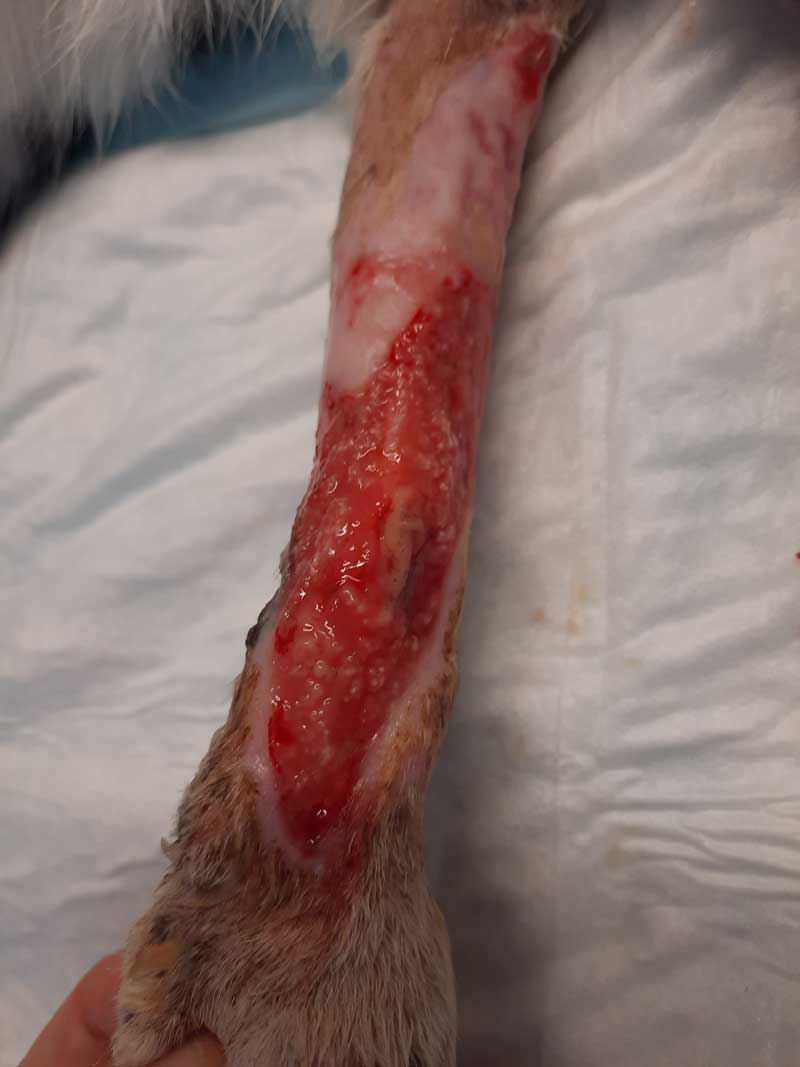
The cat was sedated to flush and redress the wounds every three days with alginate dressings covered by a sterile swab and then by a three-layer bandage (Figures 8 and 9). Although exudate continued to be profuse initially, this gradually reduced (Figure 10) and by day 26, the wounds had reduced in size by nearly half.
By day 32, the wounds had reduced further and conscious dressing changes were performed (Figure 11).
The author’s team decided to stop alginate dressings and redress with conformable adhesive dressings, which were tolerated well. By day 40, the left forelimb wound had healed well with some scar tissue.
The right forelimb wound, which had initially been smaller, had begun to over-granulate and advice once again was sought on VIN. Dressing frequency was increased to every 48 hours.
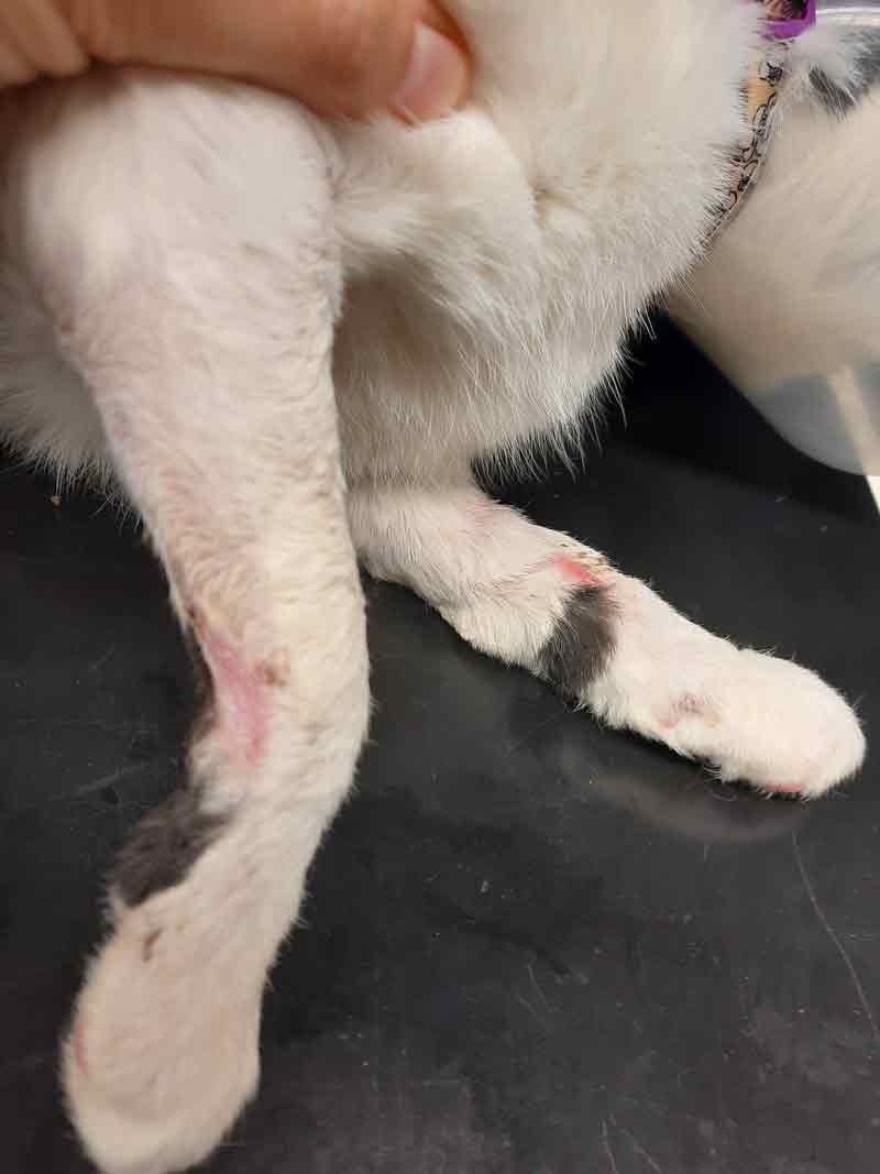
Silver sulfadiazine cream was recommended for the left forelimb to reduce over-granulation. This proved effective and both wounds had completely healed by day 50, with no further treatment required (Figures 12 and 13).
Alginate dressings, derived from kelp (O’Dwyer, 2010), are extremely absorbent (Barbu et al, 2021), retaining large amounts of water (100% to 1,000% of their dry mass; Gupta and Edwards, 2009), while providing a moist environment. The dressings conform well to the wound and can also be used in rope form to pack cavities (Wietlisbach, 2014).
Alginate dressings always require a secondary dressing to keep them in place (Gupta and Edwards, 2009). In this case, a sterile swab was used, covered by a three-layer dressing.
The alginate dressing used to treat this patient included silver, which has been shown to demonstrate efficacy against micro-organisms in vitro environments (Percival and McCarty, 2014).
The wound should be cleaned with saline and left moist. The surrounding skin should be dried. The alginate should be applied dry to the wound surface with a margin of at least 2mm around the wound edges (Leveriza-Oh and Phillips, 2012).
In the inflammatory stage, wound management was focused on removal of necrotic tissue, using mechanical debridement and wet-to-dry dressings. Subsequent manuka honey dressings provided autolytic debridement, as well as antimicrobial properties.
During the proliferative phase, silver-impregnated alginate dressings maintained, but did not disturb, the wound environment, and silver sulfadiazine cream encouraged remodelling and restoration of the normal structure.
In summary, this was an interesting case that led to some sleepless nights. Although all treatment performed was well within the reach of the average practitioner, it is important to prepare the owners for a lengthy and expensive course of treatment.
Due to the amount of care required and repeated dressing changes, healing would have been unlikely to succeed on an aggressive or exceptionally nervous cat, and euthanasia should be considered.
In this case, the patient was unusually compliant, and tolerated both hospitalisation and repeated dressing changes well.
In addition, her owners were willing and financially able to return the cat to the clinic every few days.
The author would like to thank Ardmore Veterinary Practice, the VIN, R Avery Bennett, Simon Bailey, Frances Simpson, Richard Davy and all the nursing team for their help with this case, as well as Milly and her owners for permitting publication.