10 Aug 2015
Samantha Taylor
Job TitleNutrition plays a vital role in health yet, despite inappetence being a common presenting sign of many different conditions, it is often given a low priority in the treatment of sick cats.
Early attention to nutrition may avoid complications associated with reduced calorie intake, shorten hospitalisation times and aid recovery (Chan, 2009). Assisted enteral nutrition via feeding tubes provides access to the gut to allow adequate nutritional provision as well as administration of fluid and medication.
Calculation of food requirements, monitoring of intake and use of feeding tubes are a vital part of the management of hospitalised cats. This article looks at the indications for feeding tube placement and describes the technique for placing and managing both naso-oesophageal and oesophagostomy feeding tubes.
The ramifications of poor nutrition can be severe for the feline patient. A cat’s metabolism is such that it has a high demand for dietary protein and certain essential amino acids and is unable to downregulate gluconeogenesis and proteolysis in the face of starvation (Remillard, 2002). Hence, inadequate food intake results in lean muscle breakdown. This process is exacerbated by critical illness and results in a catabolic state with multiple negative consequences for the patient. A negative energy balance can result in hypoglycaemia, hypokalaemia, immunosuppression and increased risk of infection, intestinal functional alterations and delayed wound healing.
In addition, inadequate food intake can result in hepatic lipidosis, a condition not just affecting overweight cats. In fact, the majority of systemically unwell cats will have some degree of hepatocellular fatty vacuolation (Armstrong et al, 2009). Early intervention and prompt nutritional support can aid recovery and improve outcome, leading to shorter hospitalisation times and should be considered an essential component of the treatment plan for any hospitalised cat (Brunetto et al, 2010).
Importantly, syringe feeding is not an alternative to feeding tubes and is not recommended. Very few cats tolerate this and adequate nutrition is rarely provided. At best it results in stress for the patient and at worst the creation of food aversions that can persist beyond recovery from the underlying disease. Placing a feeding tube is simple, quick and effective and should be considered for all anorexic patients.
(RER) in cats (use current bodyweight in kg)
RER (kcal/day) = bodyweight 0.75 x 70.
For cats more than 2kg RER = (30 x bodyweight) + 70
Food intake below the resting energy requirements (RER; Panel 1) for more than three days can lead to fatty infiltration of the liver (Armstrong et al, 2009) as well as impaired immune function (Freitag et al, 2000) and is used as a guide for initiation of intervention. For many cats this point is reached at home prior to presentation at the veterinary clinic – emphasising the need for prompt intervention once a cat is admitted.
Don’t delay assisted feeding while diagnostic tests are carried out – this will only prolong the period of suboptimal nutrition and could negatively affect outcome regardless of diagnosis. In addition, the following situations should prompt consideration of feeding tube placement.
Assuming the underlying disease is being managed, attention should be paid to other vital aspects of managing the inappetent cat prior to placing a feeding tube. These are critically important and listed in Panel 2.
Do not underestimate the importance of a low-stress hospital environment for cats – a species sensitive to environmental stressors, which alone can cause inappetence.
Visit www.catfriendlyclinic.org for more information.
Similarly, appetite stimulants are unlikely to be effective if pain, nausea and stress remain present. The use of appetite stimulants is not appropriate for critically ill cats as adequate food intake is rarely achieved and their use could delay placement of a feeding tube and hence adequate nutrition. They can be useful in managing food aversions, mild inappetence (for example, due to behavioural conditions) and to support cats with chronic illness (such as cats with chronic kidney disease), but are not a substitute for appropriate investigation of anorexia and the placement of a feeding tube is preferred in the majority of hospitalised cats (Agnew et al, 2014).

Naso-oesophageal and oesophagostomy tubes are used most frequently as they are straightforward to place and complications are rare. Gastrostomy tubes are indicated when longer-term nutritional support may be required and can be placed surgically or under endoscopic guidance. It is best to use the most physiological tube type – one that uses the most of the functional gastrointestinal tract. See Table 1 for a comparison of feeding tubes.
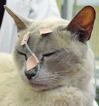
Naso-oesophageal tube placement is simple, quick and requires no sedation in the majority of cases. It is appropriate for cases where enteral-assisted feeding is likely to be required for up to one week.
Few contraindications exist, but include head trauma, absent gag reflex or impaired consciousness, uncontrolled vomiting and oesophageal and nasopharyngeal disease. Patients with nasopharyngeal disease may still benefit from this type of feeding tube, but it should be considered on an individual patient basis (for example, cat with upper respiratory tract infection requiring short-term nutrition).
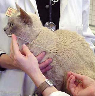
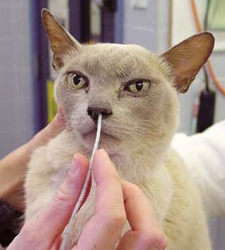
To facilitate placement, ensure you use a quiet room and gentle, minimal restraint. Bringing bedding from the cage can help the cat feel more relaxed and provide a non-slip surface. The assistant can prevent the cat moving backwards while the person placing the tube gently elevates and holds the cat’s head. The patient will most likely react to the tube passing through its nose so don’t be hesitant and, once past the nose, most cats will not react to further insertion.

Apply one vial of local anaesthetic to the nostril (usually the left nostril if right-handed) and allow one to two minutes for effect. Do not rush this stage or the nostril will not be adequately anaesthetised and the cat will react to insertion.
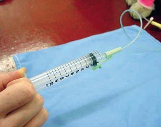
Correct placement of the tube must be checked prior to every feed to avoid complications.
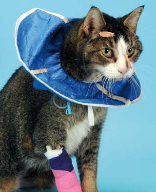
Oesophagostomy tubes are indicated when longer-term supplemented nutrition is needed (one to two weeks or more) – for example, in patients with orofacial injuries, or to facilitate medicating cats on long-term medications. If an inappetent patient is anaesthetised for another procedure, the opportunity should be taken to place an oesophagostomy tube at that time as they are easily removed if not required – for example, when performing full-thickness intestinal biopsies in a cat with suspected gastrointestinal disease.
As with naso-oesophageal tubes, persistent vomiting and oesophageal disease are contraindications as well as an absent gag reflex. As general anaesthesia is required for placement, associated risks should be considered.
The patient should be anaesthetised, an endotracheal tube secured in position, placed in right lateral recumbency and the skin on the lateral cervical area from the mandible to the thoracic inlet clipped and aseptically prepared.
Sterile gloves should be worn. Avoid cutting the tube to widen feeding holes as this can create sharp edges. Instead, stretch holes with forceps or use a blade, although this is rarely needed.
An Elizabethan/soft collar is not always needed with an oesophagostomy tube as they are generally very well tolerated. If used, a soft collar should be chosen. The stoma should be checked and cleaned at least once daily and tubes can be left in place for several weeks. To remove the tube, simply cut the suture and gently remove the tube, leaving the stoma to heal by secondary intention.
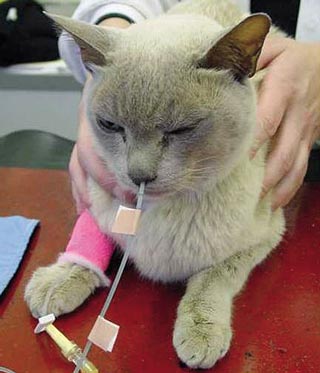
After calculation of the patient’s RER (at current body-weight) feeding should be commenced in a step-wise fashion over three days (one-third RER day one, two-thirds day two and full RER on day three). Divide the amount into four to five meals a day, or more if nausea or vomiting is observed. See Panel 3 for more information on how to use feeding tubes. Illness factors are no longer indicated due to risks of overfeeding. The choice of diet will be influenced by feeding tube type, and generally a high fat and protein diet is suitable. See Table 2 for the calorific content of various diets.
Water requirements are usually met by the liquid diet fed, intravenous fluid therapy (if used) and flushing the tube before and after use. For further information on water requirements and examples of enteral feeding charts see Korman (2013).
Feeding tubes such as naso-oesophageal and oesophagostomy tubes are inexpensive and straightforward to place, with a low rate of complications, yet they can hasten recovery from illness, shorten hospitalisation times and arrest the negative effects of malnutrition.
Any clinic hospitalising cats should prioritise nutrition as part of every treatment plan, including routine calculation of RER for all hospitalised cats and early intervention if intake falls short of this for more than three days.
Thanks go to Sarah Caney and International Cat Care who kindly supplied all the images used in this article.