5 Jun 2017
Alberto Palella Gómez presents the case of three-year-old male, neutered pug Gus in the latest Case Notes feature.
Alberto Palella Gómez
Job Title
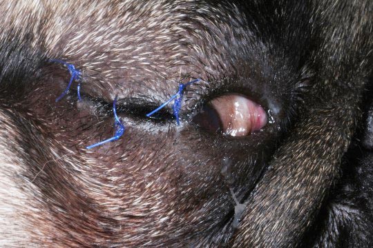
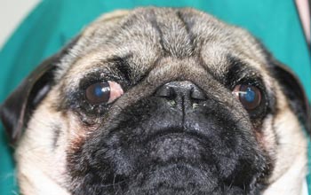
Figure 1. Gus the three-year-old male, neutered pug with lateral strabismus (exotropia) of the right eye following traumatic proptosis due to medial rectus avulsion. Other ophthalmic findings in the right eye included mydriasis, reduced corneal sensation and corneal ulceration. On the left eye, the pupil was miotic and fluorescein negative.
A case of a three-year-old male, neutered pug named Gus is presented to you one evening after being attacked by a dog in a park.
His owner has telephoned the practice reporting Gus’ right eye had prolapsed (Figure 1) and they are on their way to you.
What should you recommend regarding management of traumatic eye proptosis to the owner on the way to the practice?
The eye must be kept as clean as possible, flushing it with saline solution or tap water, if the former is not available. The owner should be advised to keep the globe moist with damp towels or clean tissue while en route to you.
After a primary and secondary survey – a detailed general examination, blood work (haematology/biochemistry) and survey x-rays, if appropriate – you have ruled out major system injuries and have stabilised your patient with appropriate analgesia, prophylactic broad-spectrum antibiotic therapy and fluid therapy.
It is time to assess the eye injury and discuss the prognosis with the owner.
What findings on a traumatic proptosis/eye injury would influence prognosis and treatment?
Traumatic proptosis is most commonly secondary to trauma and, in brachycephalic breeds, the force required may be minimal. Generally, it refers to an acute episode where the globe has been forced anteriorly beyond the rim of the bony orbit and eyelids (often becomes entrapped by these and is exacerbated by spasm of the orbicularis oculi muscle at the equator of the globe).
Strabismus as a result of torn extraocular muscles is common and the medial rectus muscle is the most frequently affected (because of its most cranial insertion). Chemosis, subconjunctival haemorrhage, corneal ulceration and uveitis are often present. Other possible findings include hyphaema, glaucoma or hypotony (in cases of scleral laceration or severe uveitis). Neuro-ophthalmic examination may reveal pupillary abnormalities and visual deficits.
Brachycephalic breeds are predisposed due to their shallow orbits, large palpebral fissures and prominent globes. On the other hand, non-brachycephalic breeds and cats are less prone to traumatic proptosis. A more severe trauma normally takes place in these cases – for example, a road traffic collision – and, thus, the prognosis for recovery of vision and/or preservation of the globe is often poor.
As a general rule, favourable prognostic indicators include proptosis in a brachycephalic dog, positive pupillary light reflexes (direct or consensual), a normal posterior segment examination and if the proptosed eye is visual. Poor prognostic indicators include proptosis in a non-brachycephalic dog, proptosis in cats, hyphaema, an obscured pupil, facial fractures, optic nerve damage and avulsion of three or more extraocular muscles (Gilger et al, 1995).
If damage to the ocular adnexae, extraocular muscles, optic nerve and/or globe is severe, prognosis for preserving even a non-functional globe is very poor and enucleation should be performed. In addition, scleral rupture might be associated with intraocular haemorrhage and loss of turgor and/or shape of the globe. In these cases, enucleation is recommended (Rampazzo et al, 2006). Transcorneal ultrasonography may assist the clinician in identifying a scleral rupture.
If repositioning is considered to be appropriate, the globe should be kept moist with ocular antibiotic ointment (for example, chloramphenicol) prior to surgery. A lateral canthotomy might be necessary in some cases, along with gentle forward traction of the eyelids to return them to a normal position. Application of a bandage contact lens during globe manipulation can help protect the corneal surface. Once the globe is replaced into its normal position, a temporary tarsorrhaphy (Figure 2) should be performed and left in situ for two to three weeks. The medial palpebral fissure can be left open to facilitate the application of topical treatments.
Topical antibiotics (for example, chloramphenicol ointment) should be used until the partial tarsorrhaphy is removed (Figure 3) and integrity of the cornea is assessed, broad-spectrum systemic antibiotics and systemic NSAIDs are also given as required.
Topical atropine sulphate 1% drops should be used with caution in cases where intraocular pressure is raised and/or tear production is reduced. However, this drug can be of benefit to reduce painful ciliary body muscle spasm and the level of secondary uveitis. Long-term complications should be discussed during the initial decision-making as well, and can include:
Always first rule out major body injuries and stabilise your patient as appropriate. The prognosis for proptosis in brachycephalic dogs can be favourable, whereas in a non-brachycephalic dog or cat, it is very poor.
Once stabilised or receiving emergency treatment, depending on the ophthalmic findings, discuss the prognosis for vision and the eye, and replace the globe as described, ensuring the owner is aware of possible long-term complications.
The author would like to thank Mike Rhodes and Rodrigo Pinheiro de Lacerda for reviewing this article and supplying images.