2 Aug 2016
Julia Smith discusses the signs and treatment options regarding this rare skin disease typically seen in older dogs.
Julia Smith
Job Title
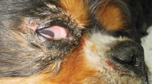
Figure 3. Erythema, crusts and alopecia of the periocular area.
Superficial necrolytic dermatitis (SND) is an uncommon skin disease in dogs. The disease is typically seen in older dogs, presents with crusting, ulcerative erythematous lesions of the foot pads, mucocutaneous junctions and genital area, and is either associated with an underlying hepatopathy or glucagonoma.
SND is also known as hepatocutaneous syndrome, necrolytic migratory erythema, diabetic dermatopathy and metabolic epidermal necrosis.
SND was first described in 1986 by Walton et al in association with diabetes mellitus in four diabetic dogs. Since then, non-diabetic dogs have been described with the disease.
The clinical features of SND bear a remarkable resemblance to necrolytic migratory erythema (NME) in humans, where distinct skin lesions observed are most commonly associated with a glucagon-secreting tumour of the alpha cells of the pancreas. SND shares many clinical (patient age, lesion distribution and morphology) and histopathologic features with NME.
The pathogenesis of SND in dogs is unknown. NME in humans is usually associated with a glucagon-secreting tumour; therefore, in humans the skin lesions are believed to be the result of hyperglucagonaemia due to a glucagon-secreting pancreatic tumour. Hyperglucagonaemia is believed to result in increased gluconeogenesis and a subsequent hypoaminoacidaemia.
Hypoaminoacidaemia develops secondarily to the catabolic gluconeogenic effects of excessive glucagon. The amino acids, histidine and lysine are essential for the continuous growth in the stratum granulosum of the epidermis. Therefore, the epidermis is susceptible to amino acid deficiency. This theory is supported by human cases in which the pancreatic tumour has been removed and the skin lesions have resolved – and the fact that with the use of somatostatin analogues, which decrease the excretion of glucagon, clinical signs have also resolved.
However, in dogs, 94% of reported cases of SND resulted from a primary hepatopathy, with only 6% resulting from a pancreatic tumour. It has been hypothesised the hepatopathy – causing decreased hepatic function – leads to a decrease in glucagon degradation (as glucagon is degraded by the liver), resulting in an increase in glucagon in the peripheral circulation.
As in humans, the excessive amounts of glucagon would result in increased gluconeogenesis and hypoaminoacidaemia, resulting in an amino acid deficiency in the epidermis. Also, due to the decreased hepatic function, there may be hypoalbuminaemia. The low levels of albumin – which carries zinc and fatty acids, essential nutrients in the epidermis – may also be partially responsible for the skin lesions. In dogs, an underlying cause for the liver dysfunction is usually not found.
Affected dogs are generally middle-aged or older with no gender or breed predisposition. SND is characterised by erosions, ulceration, crusts and exudative lesions, hyperkeratosis and hyperpigmentation of the foot pads (Figures 1 and 2), periocular (Figure 3), perioral and anal-genital regions, pressure points and mucocutaneous junctions.
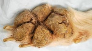
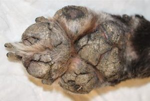
The foot pads are thickened and cracked and most of the time pain is associated with the pad lesions, often leading to lameness.
Signs of an underlying disease, such as weight loss, lethargy, polyuria and polydipsia, may also be present if there is concurrent diabetes mellitus or significant hepatic dysfunction, but, often, no signs of hepatic insufficiency are seen at the time the skin lesions develop.
It has been reported 25% to 40% of dogs with SND have concurrent diabetes mellitus. SND has also been reported in a few dogs with a history of chronic primidone or phenobarbital use as an antiepileptic (Allenspach et al, 2000).
Appropriate differential diagnoses include pemphigus foliaceus, systemic lupus erythematosus, zinc responsive dermatosis, old dog dermatosis, drug eruptions and erythema multiforme.
The diagnosis of SND is done by clinical signs, blood work, abdominal ultrasound and skin biopsies. Biopsies should be taken from recent lesions from the periphery of an ulcerating skin lesion or a crusting scaling lesion.
It is important to take multiple biopsies because the characteristic findings may not be uniformly present and biopsies from older, healing lesions may reveal only lichenification.
An elevation of liver enzymes and hypoalbuminaemia are the most common clinicopathological changes reported in the literature.
If plasma amino acid concentration is measured then there is a hypoaminoacidaemia, with most of the amino acids being less than 60% below the reference ranges. If plasma glucagon is measured, it is usually within the reference ranges or mildly increased.
In about 6% of cases with a glucagon-secreting tumour present, hyperglucagonaemia will be documented and in around 40% of cases with diabetes mellitus glucosuria and hyperglycaemia will be documented.

In dogs with SND, the abdominal ultrasound reveals the liver to be usually normal to increase in size with a mildly irregular capsule and a well-defined “honeycomb” pattern appearance to the liver parenchyma.
This pattern consists mainly of variably sized hypoechoic regions surrounded by highly echogenic borders and is pathognomonic in a dog with questionable skin lesions (Figure 4). A liver biopsy is required to confirm the unique histologic features of the hepatopathy found in SND.
Dogs with a pancreatic glucagon-producing alpha cell tumours are not reported to have the characteristic honeycomb liver changes. If this ultrasonographic pattern of the liver is not seen, evaluation for a possible pancreatic tumour is needed.

Skin biopsies of recent lesions are needed to confirm a diagnosis of SND. The histologic lesions on skin specimens are virtually pathognomonic and include diffuse parakeratotic hyperkeratosis, intracellular and extracellular oedema of the granular epithelial cells, and basal cell hyperplasia.
The histopathology reveals a classic, red, white and blue pattern. The red is parakeratotic hyperkeratosis of the superficial keratin layer, the white is vacuolated pale keratinocytes in the middle and the blue is hyperplasia of the basal cell layer, when stained with haematoxylin and eosin (Figure 5).
In dogs with an underlying liver condition, histopathology of liver biopsy samples reveals distinct regenerative nodules (seen as the hypoechoic variable-sized regions on abdominal ultrasound), bordered by severely vacuolar degeneration of hepatocytes, numerous small bile ductules, and a network of reticular and fine collagen fibres that represent remnants of collapsed hepatic lobules (the echogenic borders seen on ultrasound).
This severe lobular collapse and nodular regeneration are evidence of ongoing hepatocellular regeneration and necrosis with resultant parenchymal collapse.
Treatment of SND is symptomatic and supportive only, and has shown limited success. It is recommended dogs are placed on a high-quality, high-protein or hepatic diet, often supplemented with either cooked or raw egg yolks (three to six per day) or an amino acid powder.
Palliative therapy with parenteral and/or oral amino acid supplementation, as well as zinc, vitamin E and omega-3 fatty acid supplementation, has been reported to improve some cutaneous lesions in dogs, but for only a limited amount of time as it does not address the underlying hepatopathy.
Secondary infections must be treated appropriately, with oral or topical antibiotic and antifungal medications, with consideration to not use potential hepatotoxic drugs. Diabetes mellitus, if present, requires appropriate treatment and management.
Steroid therapy must be used cautiously and may even be contraindicated as it may predispose the dog to glucose intolerance and, eventually, diabetes mellitus.
If a glucagonoma is found, removal of the mass may lead to remission of clinical signs and increased amino acid concentrations. In humans, somatostatin analogues (a potent inhibitor of glucagon) have resulted in remission of NME lesions and shown to be potentially beneficial when a glucagonoma is present.
Resolution of clinical signs followed the removal or reduction of phenobarbital in some reported cases of SND, but other reports show no improvement with discontinuation of phenobarbital.
The prognosis for SND is poor. In one study (Miller et al, 1990), the mean survival time from the onset of skin lesions is described as 5.3 months and from the time of diagnosis as 1.6 months. Dogs are often euthanised for uncontrollable diabetes mellitus, anorexia, vomiting, diarrhoea and hepatic encephalopathy.