19 Sept 2023
Steve Butterworth shares a canine case that suffered from this condition, and its links to lameness in dogs of the same or similar breeds.
Steve Butterworth
Job Title
Pigg the cocker spaniel.
“Pigg” is a seven-year-old, male, neutered cocker spaniel weighing 17kg. He was presented to the referring veterinary surgeon in March 2022 with a history of an acute onset, severe right pectoral limb lameness after being hit by a wave while paddling in the sea.
Pain and swelling in the region of the elbow led to radiography, which revealed the presence of a lateral humeral condylar fracture. Pigg was then referred to Weighbridge Referral Centre for treatment of this injury.
On taking the history from his owner, it transpired that Pigg had shown an intermittent, mild lameness affecting that limb during the month prior to his injury.
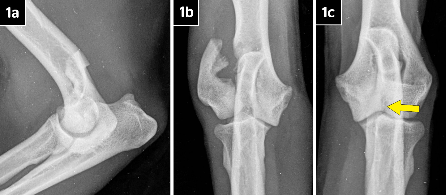
Clinical examination revealed nothing other than a 10/10 lameness affecting the right pectoral limb, associated with a painful, swollen elbow. Radiography confirmed the cause of lameness to be a lateral humeral condylar fracture (Figures 1a and 1b), but also showed subtle evidence of a humeral intracondylar fissure (HIF) affecting the left elbow (Figure 1c; yellow arrow).
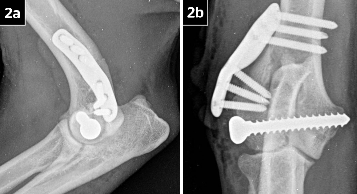
Treatment of the right humeral condylar fracture involved open reduction by way of a lateral approach to the bone.
A 4.5mm glide hole was created in the lateral condylar fragment in a retrograde fashion. The fracture surface surrounding the glide hole appeared sclerotic and a 2mm drill bit was used to create three small, shallow holes into this surface.
Once the fracture was reduced, a 3.2mm pilot hole was created in the medial fragment. This was then tapped and a 4.5mm cortical bone screw placed across the humeral condyle in lagged fashion.
A short 2.7mm lateral epicondylar anatomical plate (LEAP) was then applied to the lateral epicondyle and secured with six 2.7mm titanium locking screws. Bone that had been collected from the drill bits used to create the gliding/pilot holes in the condyle was then packed around the epicondylar fracture line.
Closure was routine and manipulation revealed a normal range of elbow joint motion, and postoperative radiographs showed good fracture alignment and implant placement (Figures 2a and 2b).
With eight weeks of restricted activity, Pigg made an excellent recovery and by that time was showing no lameness.
Radiographs taken at that stage showed good healing of the epicondylar fracture, but some evidence of an intracondylar fracture line was still present (Figure 3).
At this point, the possibility of an HIF present in the left humeral condyle was discussed with his owners, but the decision was taken to do nothing further at that time.
Although Pigg regained full use in his right pectoral limb, he then went on to develop an acute onset, severe left forelimb lameness after running into another dog in October 2022.
Radiography confirmed a lateral humeral condylar fracture (Figure 4a) and treatment was almost an exact mirror image of the previous description for the right elbow (Figure 4b).
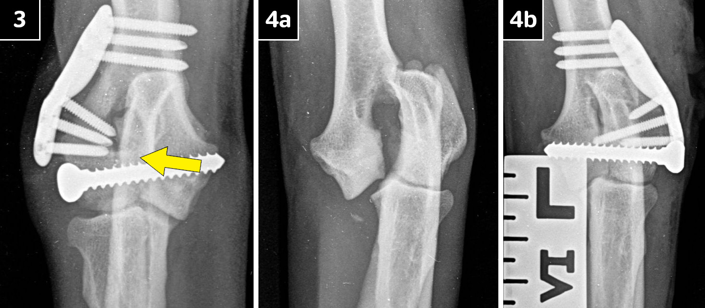
After a second eight-week period of exercise restriction, Pigg made a full recovery and radiographs showed good healing, although a small defect in the humeral condyle was noted to possibly indicate the presence of a persistent fissure (HIF).
Pigg was signed off from the surgeon’s care in December 2022 and, so far, has enjoyed a normal, unrestricted lifestyle.
Pigg’s case is of interest because of the propensity for spaniel breeds (mainly the springer in the UK1,2 and cocker in the US) to develop lameness as a result of HIF alone, or the development of humeral condylar fractures that have been predisposed to by HIF being present.
The nature of HIF is uncertain3,4, but it seems most likely that it represents a stress fracture that develops because of some form of incongruity within the elbow joint.
In Pigg’s case, the intermittent lameness seen in the right forelimb for weeks before the humerus actually fractured is suggestive of a predisposing HIF.
The suggestion of a fissure in the left condyle at the time the right was treated adds weight to this possibility, and such a problem may explain the incomplete healing of the intracondylar part of the fracture in follow-up radiographs of both elbows.
Use of CT might have helped clarify some of these points, but is not available at this centre. Little could have been done to influence the progression of Pigg’s case, except perhaps considering treating the left humerus prophylactically, before it actually fractured, by placement of a transcondylar bone screw.
The rationale for such an approach is that one study showed 18% of springer spaniels with HIF detected as an incidental finding went on to develop condylar fractures within a two-year follow-up period5.
The use of a LEAP to stabilise such fractures is a relatively recent development. The design of the plate is based on the topographical analysis of the distal humerus of several springer spaniels and, even more recently, of French bulldogs. It also takes into account the common fracture configurations to determine where best to place the plate holes.
The plates are locking and the plate holes are directed to aid achieving maximum purchase in the bone available. The plates will fit the majority of patients of these two breeds and some other similar breeds, such as cocker spaniels like Pigg.
Unfortunately, where HIF is a predisposing cause of fracture, the possibility always exists that the condylar part of the fracture will not heal fully, in which case the transcondylar implant may be subject to fatigue failure in due course, leading on to lameness resulting from implant failure or even re-fracture of the bone.
Let us hope that does not happen in Pigg’s case – he is such a lovely dog, though not very expressive (Figure 5).