6 Nov 2017
Marge Chandler discusses developments in the use of live bacteria and yeasts to combat various companion animal disorders.
Marge Chandler
Job Title
The gastrointestinal tract (GIT) is a complex ecosystem populated by tens of trillions of microbes. The number of microbial cells in mammals is 10 times that of the host cells. The microbiota, previously termed the microflora, is the microbial community on a mucosal or skin surface (Schmitz et al, 2014).
Every individual has its own unique intestinal microbiome, with variation along the GIT. The density and diversity of species increase exponentially, moving from the stomach to the colon – where the microbial content is the highest. Organisms from the phyla Actinobacteria, Bacteroides, Bifidobacterium, Firmicutes, Fusobacterium and Proteobacteria comprise most of the organisms (Jugan et al, 2017). Due to accessibility, the faecal microbiota are often analysed, although it does not necessarily reflect the rest of the GIT.
The intestinal microbiota are necessary for the development of the intestinal epithelium and the immune system. The normal microbiota play a major role in protection of the host from invasion by harmful bacteria through exclusion of potentially pathogenic organisms (such as Salmonella, enterotoxigenic Clostridium perfringens and Campylobacter species).
Suggested defence mechanisms against harmful microbes include competition for oxygen and nutrient substrates, competition for mucosal adhesion sites, creation of a restrictive environment for non-resident bacteria (such as production of substances toxic to other bacteria, changes in pH and redox potential, and hydrogen sulphide production), and secretion of antimicrobial substances (such as bacteriocins; Wynn, 2009).
The microbiome is critical for maintenance of health. “Germ-free” animals are able to survive, but are deficient in immune, gastrointestinal (GI) and, likely, brain functions. By fermenting fibre, the microbes produce short-chain fatty acids (SCFAs), including butyrate. Butyrate provides energy for enterocytes, affects the GIT barrier function, and has anti-inflammatory and anti-oxidative potential (Patel and DuPont, 2015).
In addition to the effects of the microbiome on GI disorders, evidence exists for a role in many conditions, which are not primary GI diseases, such as allergy (including canine atopic dermatitis), metabolic disease, diabetes mellitus, kidney disease, oxalate urolithiasis, periodontal disease, neoplasia and obesity. Other areas of interest in human medicine include roles of the microbiome in liver disease and hepatic encephalopathy, cognition and brain health (gut-brain axis), pancreatitis, genitourinary tract infections, and rheumatoid arthritis (Moloney et al, 2014).
The intestinal microbiome can be affected by diet (including prebiotics), antibiotics and probiotics. Large changes in macronutrient (for example, protein, fat and carbohydrate, including fibre) content can induce shifts in the composition of the microbiota (Suchadolski, 2016). Prebiotics are non-digestible foods or nutrients (such as fermentable fibres) that promote the growth or activity of beneficial bacteria. Prebiotics can cause functional changes, such as increases in butyrate (Suchadolski, 2016).
Some pet foods include prebiotics, such as inulin from chicory roots or fructooligosaccharides. Synbiotics are combinations of probiotic microbes and prebiotics provided to feed them.
Antibiotics have a very pronounced effect on the intestinal microbiome and may disrupt the microbial ecosystem for weeks to months (Suchodolski, 2016). While a full discussion is outside the scope of this article, the effect on the microbiome should be taken into consideration with the use of antibiotics.
Probiotics are live microorganisms that, when administered in adequate amounts, confer a health benefit on the host (Hill et al, 2014). Observed changes in the microbiota from probiotic use depend on the methodology used and the evaluated localisation within the gut. Generally, studies using metagenomics show probiotics can induce microbiota changes in the large intestine, but these changes typically are minor and last only a few days after the end of administration. The ability to detect an administered probiotic in the faeces of healthy dogs is also generally dose dependent. Because of this, administration of high doses over prolonged periods of time is usually required to maintain viable counts of probiotic species.
Probiotics can cause a transient increase in the administered species, but this has usually an insignificant impact on the entire intestinal microbiome. Lactobacillus species increased from 1% to 2.5% of the total bacteria after administration of a multi-species probiotic containing several different Lactobacillus species (Garcia-Mazcorro et al, 2011). A mucosa-adherent probiotic may be able to affect the microbiota more significantly; administration of VSL#3 to mice resulted in major changes in ileal microbiota (Mar et al, 2014).
Probiotic products contain different microbes, or different combinations and concentrations of them. Probiotic counts are reported in colony forming units (CFU) per dose. In humans, effective doses range from 100 million to several trillion CFU per day, depending on the particular probiotic and benefit. Levels of live probiotics can decrease while on the store shelves, so products labelled with a live content “at time of manufacture” may not be relevant and a product labelled with CFU through the end of its shelf life is more useful.
Even with those products that appear to be the same, a difference in strains can occur. This means trying one probiotic and having it work or not work does not mean we can extrapolate those results to other products or microbes. Manufacturing processes can affect the bacteria in probiotics, and it is also not possible to extrapolate the effects in one species to another, such as from dogs to cats. A potential diet effect also exists on the usefulness of a probiotic, and individuals respond differently. The best results will probably be from using a product and dose shown by research to be effective in the disorder and species being treated.
The most common research and use of probiotics so far is for GI disorders. The potential benefits of probiotics include competing with intestinal pathogenic bacteria, effects on junction stability between cells, mucus production, intestinal epithelial cell functions, mucosal immune responses and effects on the general immune system function.
Lactobacilli also synthesise some B vitamins (niacin, pantothenic acid, pyridoxine, biotin and folic acid) and some of the lipolytic (fat) and proteolytic (protein) digestive enzymes. Probiotics can potentially stimulate the innate immune response against microorganisms and dietary antigens, and can protect against pathogen-induced injury and inflammation (Erickson and Hubbard, 2000).
Acute diarrhoea has many potential aetiologies, including diet changes, infectious diseases or stress. The probiotic Enterococcus faecium SF68 may have a benefit in stress-induced diarrhoea.
Studies have shown some benefits for kittens with naturally occurring acute diarrhoea, shelter (rescue) animals with acute diarrhoea, puppies with parvovirus enteritis, and stress-related diarrhoea in sled dogs and kennelled dogs (Czarnecki–Maulden et al, 2007; Bybee et al, 2011; Arslan et al, 2012; Gore and Reynolds, 2012; Kelley et al, 2012). In dogs with acute gastroenteritis, a multi-species probiotic shortened the time to production of a normal stool from 2.2 days to 1.3 days (Hersted et al, 2010).
Bifidobacterium animalis strain AHC7, administered to dogs with acute idiopathic diarrhoea, shortened mean time to resolution of signs (Kelley et al, 2009). Supplementation of the probiotic Lactobacillus acidophilus with the prebiotic inulin to dogs decreased faecal clostridia species (Strompfová et al, 2013) and cats given L acidophilus DSM 13241 showed increased elimination of Campylobacter organisms (Baillon and Butterwick, 2003).
Dogs with diarrhoea due to distemper virus given a Lactobacillus murinus probiotic had improved clinical signs compared to those given placebo (Delucchi et al, 2017). Stool consistency and amount, appetite and mental status were better in treated dogs, although vomiting was not improved.
Decreased appetite, food aversion, vomiting and diarrhoea are common in pets given antibiotics. Antibiotics also disrupt the GI microbiome, causing a dysbiosis.
A multi-strain synbiotic used in cats given clindamycin showed the treated cats were more likely to have completed the treatment due to less vomiting. The synbiotic-treated cats also had better appetites (Whittemore et al, 2016). The yeast Saccharomyces boulardii shortened the length of time diarrhoea was present in dogs given lincomycin and prevented diarrhoea when given concurrently with it (Atkas et al, 2007).
Dogs with diarrhoea and inflammatory bowel disease (IBD) generally show less intestinal microbial diversity than normal dogs (Figure 1).
Dogs with IBD supplemented with E faecium had significantly increased richness of the faecal bacterial microbiome, which became more similar to that of healthy dogs (Schmitz et al, 2014). Probiotic supplementation to 21 dogs with food-responsive diarrhoea showed beneficial effects on intestinal cytokines and microbiota (Sauter et al, 2006), although the changes were not significantly associated with the clinical response, as the dogs responded mostly to a hydrolysed diet.
A probiotic containing multiple bacteria given to 10 dogs with long-term IBD resulted in significant decreases in the Canine IBD Activity Index, intestinal histology scores and decreased intestinal infiltration with CD3+ T-lymphocytes (Sauter et al, 2006).
Another study on the effects of VSL#3 compared to treatment with prednisolone and metronidazole in 20 dogs with IBD showed a significant decrease in clinical and histological scores with both treatments (Rossi et al, 2014). The dogs treated with probiotics also had a normalisation of the microbiome dysbiosis.
Fewer studies have been reported on the use of probiotics in cats with chronic diarrhoea. Use of a synbiotic in adult cats with chronic diarrhoea resulted in an improvement in stool quality (Hart et al, 2012). Administration of Lactobacillus group 2 and E faecium to 27 juvenile cheetahs was associated with a significantly increased bodyweight and improved faecal quality compared to controls (Koeppel, 2006).
Oxalate is eliminated through urinary excretion, forming insoluble calcium oxalate in the GIT with faecal elimination or by oxalate degradation by GI microorganisms.
In humans, some probiotics containing Lactobacillus species or Oxalobacter formigenes degrade oxalate in the intestine, resulting in decreased absorption and decreased urinary excretion (Lieske et al, 2005). Dogs with O formigenes in their faeces appear to have a lower risk of calcium oxalate urolith formation. The prevalence of O formigenes in faecal samples from dogs with calcium oxalate uroliths was 25%, compared to 50% in healthy dogs and 75% in healthy dogs of breeds not at risk for oxalate uroliths (Gnanandarajah et al, 2012).
A study of the lactic acid bacteria in dog and cat faeces suggests manipulation of GI bacterial components may decrease intestinal oxalate, similarly potentially decreasing intestinal oxalate absorption and renal excretion (Weese et al, 2004). The faeces of 86% of healthy cats has the genes for O formigenes, although the association with oxalate urolithiasis has not yet been explored (Weese et al, 2009).
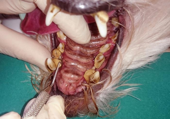
The dental biofilm – a microbial community attached to the tooth surface – is a main cause of dental pathology (Figure 2). A mixed culture probiotic with Lactobacillus acidophilus LA-5 and Bifidobacterium bifidum BB-12 was shown to have an in vitro bactericidal effect on pathogenic species from the supragingival sites of dogs with dental disease (Zambori et al, 2016). This suggests the antimicrobial effect of the probiotics may be useful in the prevention and control of dental plaque, the cause of periodontitis.
Nitric oxide (NO), an important inflammatory mediator, is increased in human periodontitis and agents blocking the production of NO or its effects might be therapeutically valuable. The probiotic L brevis contains high levels of arginine deiminase, which inhibits NO generation by competing with NO synthase for arginine. In humans, topical application of L brevis decreased inflammatory mediators involved in periodontitis. Preliminary results of topical L brevis CD2 in dogs showed reduced gingival inflammatory infiltrates (Vullo, 2014).
The gut microbiota differs between obese and lean individuals. The bacterial groups and subgroups (Firmicutes and Bacteroidetes) comprise 90% of the intestinal bacteria.
Some studies report decreased proportions of Bacteroidetes and sometimes an increased proportion of Firmicutes in obese humans. A gut microbial composition favouring increased Firmicutes may cause more efficiency in energy extraction from the diet, resulting in higher levels of SCFAs. These SCFAs are suspected of altering the metabolism of obese individuals and can provide up to 10% of the daily energy supply (Million et al, 2013).
An increase in faecal proportions of Bacteroidetes and decrease in Firmicutes during weight loss has been reported (Arora et al, 2013). Compared to lean beagle dogs, obese dogs were found to have less intestinal microbiota diversity. Proteobacteria was the predominant bacteria in obese dogs and, unlike humans, Firmicutes are predominant in lean dogs (Park et al, 2015). It is possible the maternal diet and early gut colonisation may both play a role in the pathogenesis of obesity (Kaplan and Walker, 2012).

One small study looked at the effects of a probiotic as a therapy for feline obesity. Short-term use of E faecium SF68 dietary supplementation in eight cats had no significant effect on food intake, bodyweight, body composition or metabolic parameters in overweight and obese cats (Kathrani et al, 2016; Figure 3). Further studies should examine this concept, as obesity is a significant medical problem in pets.
In humans with stage 3 and 4 chronic kidney disease, a decrease in azotaemia was seen in patients with six months of probiotic treatment (Ranganathan et al, 2009). Similarly, in azotaemic cats, the serum urea nitrogen and creatinine concentrations decreased after 60 days’ administration of a probiotic; although the concurrent treatments varied and the relationship to quality of life and survival time was not clear (Palmquist, 2006).
In contrast, in another study in cats with naturally occurring azotaemia, supplementation with a synbiotic had no effect on the level of azotaemia (Rishnew and Wynn, 2011).
The skin has a microbiota population of bacteria and fungi. The mycobiota (fungal population) of dogs’ allergic skin is significantly less rich and diverse than that of healthy skin (Meason-Smith et al, 2015).
Humans and dogs with atopic dermatitis (AD) have a skin microbiota dysbiosis, with lower diversity of microbial populations than healthy individuals (Rodrigues Hoffmann et al, 2014). Whether altered microbial populations are the cause or the effect of inflammatory skin conditions is unknown (Rodrigues Hoffman, 2017). Some evidence exists that AD can be prevented in humans with the use of probiotics, especially if administered during early infancy (Kim and Ji, 2012).
Studies on the prevention or treatment of canine AD have had mixed results (Marsella 2009; Kim et al, 2015). One study showed a decrease in the use of prednisolone in dogs with mild to moderate AD given Lactobacillus paracasei K71 compared to those on cetirizine hydrochloride (Ohshima-Terada et al, 2015).
Where possible, choose a probiotic product that has been tested for the disorder and in the species being treated. Individuals within a species also have different microbiome composition, which will affect how the probiotics work.
Using a probiotic for acute gastroenteritis may shorten the number of days of diarrhoea, although these cases may also be self-limiting. Where a stressful event is predicted, using a probiotic for several days prior to and during the event may be useful. Probiotics may have a beneficial role in chronic enteropathies as adjunct therapy.
The potential for use in weight management, calcium oxalate urolithiasis prevention and dental disease is encouraging, although studies have not yet provided enough information for recommendations.