9 Dec 2019
James Colver discusses a potentially life-threatening – and under-reported – complication seen with endotracheal intubation.
James Colver
Job Title
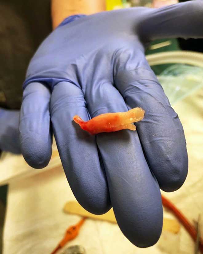
Figure 1. The obstructive fibrinous tracheal pseudomembrane was removed in one piece.
Endotracheal intubation is a common procedure in veterinary medicine. It provides a means of establishing a patent airway and, following inflation of the cuff, provides a leak-free connection of the patient to the anaesthetic circuit – enabling precise delivery of anaesthetic agent and a means of providing positive pressure ventilation.
The use of endotracheal tubes (ETTs) with inflatable cuffs also protects the patient from aspiration of gastric contents or material entering the oropharynx, and protects staff from environmental exposure to anaesthetic gases. A significant number of complications are associated with endotracheal intubation, and it has been associated with increased morbidity – particularly in cats1-3.
Complications can range from minor to life-threatening, including inadvertent endobronchial intubation, occlusion of the tube with foreign material or patient secretions, ischaemic injury to the tracheal mucosa, tracheal stenosis, tracheal rupture, damage to the larynx and arytenoids, the patient biting and inhaling a broken part of the tube, and difficulty in removing the tube postoperatively3-7. Obstructive fibrinous tracheal pseudomembrane (OFTP) formation is a rare, but potentially fatal, complication of endotracheal intubation, and has been reported in humans8-11 and at least one dog12.
A previously healthy three-year-old female Boston terrier with a body condition score of 4/9 presented with dystocia that was deemed surgical. She was induced for a caesarian section, and intubated with a 6.5mm red rubber ETT. The cuff was inflated with 3ml of air using the leak-test method. Gastroesophageal reflux was noted on intubation.
The general anaesthetic lasted for two hours, and was unremarkable, except for a period of hypercapnia that resolved after she was mechanically ventilated.
On return of the swallowing reflex and deflation of the ETT cuff, extubation was not possible, as the ETT appeared to be stuck in the trachea. The dog was reinduced and given an IV dose of 0.06mg/kg dexamethasone. Lidocaine, together with a sterile, water-based lubricant, was instilled around the ETT, and after 45 minutes it was removed successfully.
Further investigations were postponed due to the risk of re-anaesthetising the dog, but her recovery was uneventful and she was discharged later that day. She re-presented within 12 hours with dyspnoea, stertor and cyanosis, and a tracheostomy was performed.
The tracheostomy tube was removed the following day and the dog appeared to be coping well for a further 24 hours, at which point she became dyspnoeic again, necessitating another tracheostomy tube. Repeated attempts were made over the following four days to remove the tube, but each time her breathing rapidly deteriorated. Radiography revealed interfascial emphysema in the neck and poor definition of the trachea. She was anaesthetised to allow endoscopic examination of the upper airway.
A greyish mass of what appeared to be mucus was documented just caudal to the larynx, occupying almost the entire tracheal lumen. An attempt was made to clear the plug, firstly by using an endoscopic cytology brush, and then by grasping it with endoscopic biopsy forceps. Neither attempt relieved the obstruction. A pair of alligator forceps was then directed under endoscopic guidance to grasp what was now recognised as solid tissue.
Although the mass could be firmly grasped with the forceps, only small fragments of the tissue could be broken off, and the plug remained firmly fixed in the trachea. It was noted the forceps and the endoscope could be passed alongside the mass to the extent it was possible to visualise the tracheostomy site caudally to the obstruction. At this point, a non-cuffed ETT of the same diameter as the trachea was passed through the larynx and rotated as it was passed into the trachea, in the hope it may break down what was adhering the mass to the tracheal wall.
The ETT was retracted and the mass successfully removed in one piece. On examination, it resembled a silicone mould of the trachea, and was firm and rubbery in texture (Figure 1). The trachea was re-examined to confirm the blockage had been successfully removed. On the dorsal wall of the trachea just cranial to the tracheostomy was a 2mm by 3mm patch of what appeared to be granulation tissue. The rest of the tracheal mucosa was normal in appearance.
The dog recovered well, but within 24 hours had started to develop respiratory distress again. During an attempt to replace the tracheostomy tube, she expectorated another tracheal cast, after which she demonstrated a drastic improvement, and no subsequent relapses were noted.
Three days later, her tracheostomy stoma had almost healed, and she was breathing appropriately through her own airway.
OFTP has been attributed as a cause of failed extubation in humans10, although in the case described here it is unclear whether the membrane formation was a cause or consequence of the difficult extubation.
Mitigating the risks associated with endotracheal intubation requires an understanding of the different tube types available, the techniques recommended to intubate patients, and an ability to recognise and respond to problems without delay.
ETTs are manufactured from materials including polyvinyl chloride (PVC), silicone and red rubber. PVC tubes are made with a preformed curve to facilitate visualisation of the larynx on intubation. Silicone tubes are straight and softer than PVC or rubber, and sometimes require the use of a stylet for intubation – particularly in the smaller sizes and in cases where visibility is limited. Both PVC and silicone tubes are able to conform to the shape of the trachea as they warm up7.
Red rubber tubes are opaque, making it impossible to visualise luminal obstructions. The rubber perishes and cracks over time, making effective cleaning and disinfection impracticable. They feature a high-pressure, low-volume cuff that provides superior protection of the airway, but is more likely to damage the tracheal mucosa than the low-pressure, high volume cuffs4. Red rubber tubes have fallen out of favour in veterinary medicine7, and it has been suggested their use in dogs and cats should be discontinued13.
Various techniques have been described to adequately inflate the cuff without causing tracheal damage. The simplest involve inflating the cuff to a predetermined degree of inflation of the pilot balloon, or for one person to squeeze the reservoir bag on the anaesthetic circuit with the valve closed while another person inflates the cuff, listening at the mouth until leaking around the tube can no longer be heard. These techniques are highly subjective and have been shown to result in excessive pressure in the cuff14-16,18. The safest method of inflating the cuff is with a pressure gauge15. In humans, the recommendation is to inflate the cuff to between 20cm to 30cm H2016. Safe cuff pressure for dogs and cats has been suggested between 19cm to 24cm H2O4,13,14,17, although no absolute consensus exists on a maximum safe value exists.
OFTP is an intraluminal cast encircling, and attached to, the tracheal wall consisting of fibrin, inflammatory cells and desquamated necrotic epithelium. OFTP cases have been reported in humans at the site of the cuff and are primarily thought to be a result of ischaemic injury to the tracheal mucosa, secondary to high pressure in the cuff8, although the exact mechanism behind their formation remains unclear. One case of OFTP has been described in a human where cuff pressure was maintained at 15cm H2O9, but the patient had a history of vomiting and a difficult intubation, suggesting caustic injury as an aetiological factor.
Hypoperfusion secondary to hypotension has been implicated as a cause of ischaemic insult to the tracheal mucosa and subsequent OFTP formation19. Other risk factors have been proposed, including inappropriately large ETT, traumatic intubation, and the length of time a patient is intubated11. Diphtheria infection is also associated with pseudomembrane formation in respiratory tracts9.
One case of OFTP has previously been described in a dog12, with a presentation almost identical to this case. The pseudomembranes in both dogs were observed at the site of the cuff, both dogs presented with respiratory distress at a similar time following extubation, and the pseudomembranes from both were histologically similar.
The characteristics of both presentations are supportive of tracheal injury secondary to the cuff. Additionally, both dogs were brachycephalic – breeds with a higher risk of gastroesophageal reflux during general anaesthesia. Interestingly, both cases had an OFTP recurrence, which differs from many described in human medicine.
To conclude, OFTP is a rare and potentially fatal complication associated with endotracheal intubation and requires urgent intervention. It should be considered in any unexplained occurrence of respiratory failure, stertor or suspicion of upper airway obstruction after intubation. Immediate endoscopic OFTP removal is recommended after the patient has been stabilised with oxygen, and in recurring cases, corticosteroid therapy, together with inhaled unfractionated heparin and aerosolised N-acetylcysteine, has also been suggested12.
The recurring nature of the cases described to date indicate it may be prudent to hospitalise patients for at least two days following retrieval.