5 May 2023
Erinn Mills BSc, DVM, Andrew Lewin BVM&S, DACVO and Renee Carter DVM, DACVO cover points for consideration when deciding to pursue this treatment.

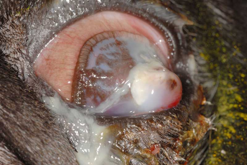
Figure 1. A colour photo of a canine patient with chronic primary glaucoma of the left eye. The eye is buphthalmic with ocular discharge, episcleral injection, diffuse corneal oedema and multifocal Haab’s striae. Lens subluxation and a superior aphakic crescent are also present. This eye was non-visual and despite medical management, frequent increases in intraocular pressure occurred. Enucleation was recommended.
Many factors are involved in making an informed decision to perform an ocular salvage procedure – such as enucleation – in canine and feline patients.
While the decision is straightforward in many cases, clinicians may occasionally wonder if performing a salvage procedure is the best option. Salvage procedures are generally reserved for situations when serious ocular disease results in permanent blindness and pain.
However, other factors may influence this decision, such as concern for neoplasia or owner/patient compliance.
In this article, the authors will outline several factors that can be considered by clinicians faced with making this decision.
In most circumstances (with the exception of neoplasia), salvage procedures are reserved for eyes considered likely to be permanently blind.
Vision in mature cats and dogs can be readily assessed using the menace response, visual tracking, maze testing or visual placement tests. The clinician should carefully consider whether vision loss is likely to be temporary or permanent; for example, an acute intraocular pressure elevation in a glaucomatous eye may cause potentially reversible temporary blindness.
Many dogs are permanently blind in one or both eyes, but do not necessarily require an ocular salvage procedure. Only when vision loss is accompanied by ocular disease resulting in severe or chronic pain is a salvage procedure likely to be necessary. For example, patients with extensive cataracts frequently have significant visual impairment, but are unlikely to suffer from discomfort as long as secondary intraocular inflammation is appropriately managed.
Similarly, animals with primary retinal disorders (for example, progressive retinal atrophy or sudden acquired retinal degeneration syndrome) are likely to have significant visual impairment, but are unlikely to suffer from any ocular discomfort if appropriately managed.
If ocular pain is likely to be severe, persistent/recurring, and challenging to control, a salvage procedure should be considered.
Commonly encountered ocular conditions associated with severe persistent pain and loss of vision include end-stage glaucoma (Figure 1), endophthalmitis and full‑thickness corneal defects (Figure 2). Conditions such as glaucoma frequently result in chronic ocular pain that goes unnoticed by owners.
Dogs with glaucoma appear to have an increased expression of “normal” behaviour after enucleation (Bujan et al, 2021). Following enucleation for chronic glaucoma, sensitivity to mechanical stimulus both locally (around the orbit) and at remote sites (metacarpus and metatarsus) decreases (Zibura et al, 2021).
Multiple studies have shown that most owners are very satisfied with their pet’s quality of life after it undergoes unilateral or bilateral enucleation (Hamzianpour et al, 2019; Palmer et al, 2021).
Primary intraocular neoplasia may warrant a salvage procedure in certain situations. Clinical signs of intraocular neoplasia may include the presence of a visible mass, dyscoria (abnormal pupil shape), hyphaema, uveitis, or secondary glaucoma.
Histopathology should be completed following globe removal as this is likely to inform prognosis. Common primary intraocular tumours of cats and dogs include melanoma/melanocytoma, adenocarcinoma/adenoma, feline diffuse iris melanoma and feline intraocular primary sarcoma.
Surgical management of retrobulbar extraocular disease (including neoplasia) may necessitate a simultaneous salvage procedure (for example, exenteration) in some cases.
It should be noted, however, that globe-sparing orbitotomy is possible in selected cases.
Successful medical management of potentially painful, vision-threatening ocular diseases usually requires frequently administered long-term therapy.
Some owners are unable (for example, due to financial or time constraints) or unwilling to administer such treatment protocols to their pets. In such cases, enucleation may represent a more realistic option to alleviate patient discomfort.
Patient temperament may lead the clinician to consider an ocular salvage procedure over medical management if it is likely the patient will not accept medication administration to maintain ocular comfort.
While this is not a common reason for an ocular salvage procedure to be performed, this factor should be carefully considered by the clinician in each case.
Several ocular salvage procedures are available – ranging from relatively straightforward with a low risk of complications (for example, enucleation), to more technically demanding with a higher risk of complications (for example, evisceration and intrascleral prosthesis). The choice of technique should be dictated by clinician experience level and the individual characteristics of each case.
Enucleation is the removal of the globe, conjunctiva, eyelid margins and third eyelid.
Transpalpebral and transconjunctival approaches are possible in cats and dogs, with the transpalpebral approach being the most suitable for removal of eyes affected with corneoconjunctival infections or neoplasia.
Postoperative complication rate for enucleation has ranged from 4 per cent to 6.5 per cent in recent studies (Hamzianpour et al, 2019; Palmer et al, 2021). These complications include infection, fistula formation, haemorrhage, orbital emphysema (particularly in brachycephalic breeds), cyst formation and dehiscence. Some clinicians may choose to place silicone intraorbital implants at the time of surgical closure, which may improve cosmetic appearance in some animals.
Editor’s Note: The below procedure is considered cosmetic by the RCVS and so should not be offered as an option in the UK.
Evisceration is the removal of the intraocular contents of the globe, leaving only the cornea and sclera of the globe in situ. The intraocular contents are then replaced by a silicone sphere, which is implanted prior to corneoscleral shell closure.
The primary advantage of this approach over enucleation is the preservation of ocular tissue that has the outward appearance of an intact globe. It should be noted, however, that cosmetic outcomes do vary, and certain animals will require long-term topical medication to maintain corneoscleral shell health.
Animals with pre-existing corneal disease, septic endophthalmitis or intraocular neoplasia are generally considered poor candidates for this surgery. Potential complications – which are reported to occur in 9 per cent to 16 per cent of patients – include the development of keratoconjunctivitis sicca (dry eye), scleral wound dehiscence with extrusion of the implant, postoperative keratitis and secondary entropion (Naranjo and Dubielzig, 2014).
This procedure is generally not recommended in cats as malignant neoplasia has been linked to implant failure in this species (Naranjo and Dubielzig, 2014).
Exenteration is the removal of the globe, eyelid margins and all orbital contents. This salvage procedure should be reserved to treat severe orbital disease, such as an orbital abscess, granuloma, sialocele/mucocele, or neoplasia that extends into the orbit.
The risks of exenteration are similar to that of enucleation, except a higher risk of intraoperative haemorrhage exists due to the likelihood of encountering additional vasculature. As noted previously, globe-sparing surgical approaches to orbital disease are possible in selected cases.
Ciliary body chemical ablation is a salvage procedure in which epitheliotoxic agents are directly injected into the vitreous to reduce aqueous production by the ciliary body. This procedure is most commonly performed using gentamicin, but cidofovir can also be used.
The primary indication for this approach is for reduction of intraocular pressure in glaucomatous, non-visual eyes, where enucleation either cannot be performed due to anaesthesia patient safety concerns or owner reluctance.
It should be noted, however, that post‑procedural cosmetic appearance can vary. In earlier studies, pharmacological ciliary body ablation was successful in 65 per cent of patients; however, a more recent report had a success rate of 86 per cent for the first injection (Gelatt et al, 2013; Rankin et al, 2016).
This procedure is generally not recommended in cats due to the potential for intraocular sarcoma development; post-procedure sarcoma development has also been reported in dogs (Duke et al, 2013a; 2013b).
Ciliary body ablation is contraindicated in cases where intraocular neoplasia is suspected or in patients with significant renal disease when using gentamicin for the procedure.
In conclusion, the decision to perform an ocular salvage procedure must be made on a case‑by‑case basis. Vision, pain, the presence of neoplasia and owner/patient compliance must all be simultaneously evaluated.
While enucleation is the most commonly performed ocular salvage procedure in most settings, other options are available that may represent viable alternatives for certain cases.
When uncertain regarding diagnosis, prognosis or which surgical approach is most appropriate, it is strongly recommended that specialist advice is sought.