27 May 2019
Georgina Harris presents the case of a four-year-old, neutered dachshund that presented with a 24-hour history of acute onset, progressive paraparesis.
Georgina Harris
Job Title
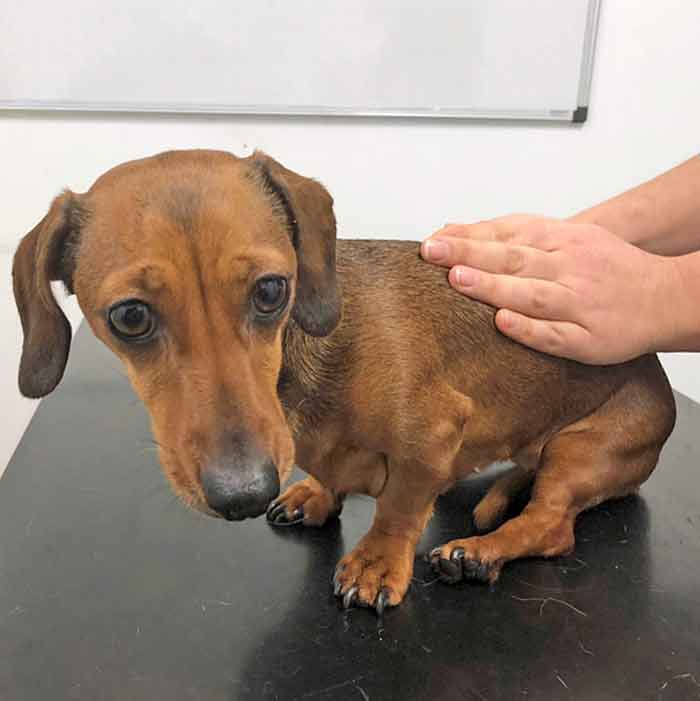
The patient exhibiting delayed paw positioning in his left pelvic limb.
Sausage – a four-year-old, male, neutered dachshund – presented with a 24-hour history of acute onset, progressive paraparesis.
A neurological examination revealed the dog was non-ambulatory paraparetic. His spinal reflexes were intact in all four limbs, with a cutaneous trunci cut off at L2. His paw positioning was reduced to absent in both pelvic limbs and his cranial nerves were unremarkable. Pain was perceived on palpation of his thoracolumbar region and his bladder was large on abdominal palpation. Deep pain perception was not assessed as some voluntary movement was retained.
From your neurological examination you localised him to a T3-L3 myelopathy. Radiographs of his spine revealed a narrowed disc space at T13-L1 and calcified disc material could be seen within the canal at this level. You are strongly suspicious of an intervertebral disc extrusion; however, the owners have declined referral for further investigation.
What is the prognosis for managing the dog conservatively, and what are the important management factors?
If your suspicion of an intervertebral disc extrusion is correct, Sausage has a good prognosis for return to ambulation with medical management alone, as he has retained deep pain perception in his pelvic limbs. A review of the literature reports a prognosis of return to ambulation at 86%, compared with surgical management at 90%. However, this is very different when concurrent loss of deep pain perception exists, when the prognosis for medical management may fall to as low as 6%, and any potential recovery is likely to be prolonged and require intensive nursing care1.
Cage rest, analgesia, bladder management and physiotherapy are the basis of medical management for an intervertebral disc extrusion. Sausage was kept in the practice on strict cage rest for a week to help prevent further extrusion of disc material and potentially allow healing of the annulus. He was monitored with twice-daily neurological examinations to assess for any progression or improvement in his neurological status.
The patient was managed initially on methadone (0.2mg/kg IV every 4 hours), meloxicam (0.05mg/kg orally once a day) and gabapentin (10mg/kg orally three times a day). He was pain scored every 4 hours for the first 24 hours. Methadone was discontinued after 24 hours and buprenorphine (0.02mg/kg IV every 8 hours) was given for the following 48 hours, as his pain was well controlled.
The dog was monitored every 4 hours for his ability to turn himself, and while he was unable to do so, his recumbency was changed to prevent the development of pressure sores. He was monitored closely for signs of bladder dysfunction as urinary retention is common in dogs that are non-ambulatory.
If any doubt existed over his bladder size this was checked with an ultrasound probe. He was carried outside on to grass regularly to give him the opportunity to urinate and assess his ambulation. Options for managing non-ambulatory dog bladders include intermittent and indwelling catheterisation, as well as manual expression.
Dogs that are non-ambulatory following intervertebral disc extrusion are predisposed to developing urinary tract infection (UTI)2, therefore a urine sample was taken by cystocentesis to check for signs of UTI. Sausage’s urine was found to be unremarkable. He also underwent gentle physiotherapy three times a day, which involved massage of his pelvic limbs and passive range of motion. Sausage responded well to medical management; he was ambulatory with light sling support and able to urinate voluntarily within one week of hospitalisation.
At discharge, the dog’s owners were advised to continue strict cage rest for four weeks and continue to monitor him for signs of pain, UTI or deterioration of neurological signs. He was sent home with five days’ gabapentin and meloxicam at the previously stated dose. The risk of recurrence of intervertebral disc extrusion was also discussed and a re-examination arranged for four weeks later.