2 Nov 2015
Claire Bradley
Job Title
Figure 1. Tonometry is a quick and easy method of measuring intraocular pressure.
Glaucoma is a severe neurodegenerative disease and represents a range of disorders. Any “glaucoma” leads to optic nerve and retinal pathology, resulting in irreversible vision loss. Common in cats, its insidious nature makes this a difficult disease to identify early.
This article aims to remind practitioners of the initial examination, diagnosis and management of glaucoma.
Normal feline intraocular pressure (IOP) varies with the time of day, age of the patient and reproductive status.
One study found normal IOP values of 19.7+/-5.6mmHg using the Tono-Pen. It was noted the Tono-Pen significantly underestimated values repeatedly, but predictably3.
One reading, obtained in a single consult, may not accurately reflect the degree of IOP fluctuation throughout the day. In addition, the type of tonometer can vary results – thus it is important to record which instrument was used and be consistent for repeat visits (Figure 1).
The examination for cats is similar to that described for dogs. Cranial nerve assessment, Schirmer’s tear test readings, IOP measurement and thorough examination of the adnexa, cornea, iris, lens and fundus should be performed. Direct and indirect ophthalmoscopies are recommended.
On fundoscopy (Figure 2), obvious species differences exist – retinal arteries are less tortuous in the cat versus the dog and retinal veins stop at the edge of the optic disc. The optic nerve head, in the non-tapetal fundus, is unmyelinated. This makes distinction of optic nerve cupping in glaucomatous eyes harder to appreciate in the cat.

Individual fibres of the pectinate ligament are very fine and relatively sparse in the cat. This, combined with a relatively deep anterior chamber and a wide irido-corneal angle, helps reduce the incidence of primary glaucoma in cats.
Although rare, cases may occur in any breed. The Siamese and Burmese breeds are slightly more predisposed6,7.
Most feline glaucoma cases are secondary to an underlying process, such as chronic lymphocytic-plasmacytic uveitis, aqueous humour misdirection syndrome, neoplasia, intraocular haemorrhage or trauma.
Pain, corneal oedema and episcleral injection are frequently associated with canine glaucoma. These are less pronounced in cats, with many owners unable to recognise their pet has an ocular problem or is in pain.
Due to its insidious nature, cats are frequently presented for blindness and/or a change in the eye’s appearance (for example, mydriasis, buphthalmos, fibrin clots and obvious intraocular masses). A retrospective study found 73% of cats with glaucoma were blind when presented8.
Haab’s striae are possible, but seldom seen, in cats with chronic glaucomatous eyes.
Uveitis is arguably one of the most frequent ocular clinical signs prompting veterinary attention, so clinicians should be aware of the risks associated with feline glaucoma that may result from untreated intraocular inflammation.
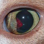
Clinical signs in cats include miosis, rubeosis iridis, keratic precipitates, hypopyon and hyphaema (Figure 3). Aqueous flare, when present, is pathognomonic for uveitis, but its absence does not rule out diagnosis.
Any of these signs should alert the clinician to the possibility of glaucoma and instigate rigorous IOP monitoring.
Anterior uveitis may lead to glaucoma due to inflammatory cells in the iridocorneal angle and ciliary cleft blocking aqueous humour outflow. Formation of anterior synechiae and/or pre-iridal fibrovascular membranes further reduce the drainage angle. Most of the feline drainage angle can be seen without a goniolens – using focal illumination and magnification.

Feline aqueous humour misdirection syndrome (AHMS) is a recognised cause of glaucoma in cats.
In this process, aqueous humour passes posteriorly into the vitreous via small breaks in the hyaloid membrane. This leads to higher vitreal pressure, anterior displacement of the lens and a resultant shallowing of the anterior chamber (Figure 4).
Clinically, AHMS is characterised by a shallow anterior chamber, intact lens zonules and a narrowed approach to an open irido-corneal angle.
Studies have found the mean age of affected cats is 11.7 years (range 4 to 16 years), with female cats more likely to be affected9.
Slit lamp biomicroscopy and ultrasonography are useful aids to diagnose this condition. On histological examination, a thickened anterior vitreal face is seen with partial ciliary cleft collapse and cavitated vitreal regions.
Surgical and medical management have been described for AHMS treatment, although a consensus on which modality is more appropriate has yet to be determined. Medical therapy aims to reduce aqueous humour production, using carbonic anhydrase inhibitors (CAI), while surgical options include lensectomy and anterior or pars plana vitrectomy.
Trauma to the lens caused by a penetrating wound (for example, cat claw injury) should always be investigated – especially in young felines. Practitioners must assess the lens as phacoclastic uveitis may lead to higher IOP.
Primary lens luxation, with secondary glaucoma, is extremely uncommon in the cat. In contrast with the dog, luxation usually occurs secondary to trauma or chronic inflammation and resultant disruption of the zonular fibres securing the lens.
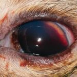
Older cats may develop hypertension causing retinal haemorrhages and hyphaema, the latter of which can lead to higher IOP (Figure 5).
Ocular neoplasia is common in cats, with iris melanomas, post-traumatic sarcomas, iridociliary adenocarcinomas and lymphoma most frequent10. Neoplastic masses may lead to higher IOP, via occlusion of the drainage angle.
In cases with raised IOP and an obvious intraocular mass, enucleation and histopathology may be required for definitive diagnosis (Figure 6).
In other cases, where the intraocular mass is suspected to be secondary rather than a primary isolated ocular mass (for example, lymphoma), additional screening tests should be performed to rule out metastatic disease. Such tests include haematology and comprehensive biochemistry, fine-needle aspiration of lymph nodes for cytology and further diagnostic imaging (thoracic radiographs, abdominal ultrasonography and/or computed tomography).
Ocular lymphoma may be successfully diagnosed on aqueocentesis, but the use of aqueous humour cytology for diagnosing other forms of intraocular tumours remains questionable. In patients with an ocular mass confirmed as lymphoma, further staging is still indicated to confirm whether the mass represents a primary or secondary lesion.
Where enucleation is performed, the affected eye should be sent for histopathological analysis. This enables an accurate diagnosis, removes the painful eye and may provide some prognostic indication.

Due to its late presentation, successful treatment is frequently unrewarding. Treating any underlying cause is more important.
Uveitis cases should receive a full work-up to determine the aetiology. Diagnostics should include a minimum database of haematology and comprehensive biochemistry, urinalysis, thoracic radiographs, abdominal ultrasound and PCR for Toxoplasma, Neospora and Bartonella. Occasionally, aqueocentesis may also be indicated.
Differentials for feline uveitis include inflammatory, infectious, immune-mediated, traumatic and idiopathic causes.
Treatment of feline glaucoma is similar to that recommended for canine glaucoma, but there are variations. Initial treatment should be aimed at the primary underlying cause. Uveitis should be treated aggressively with topical and/or systemic steroids, and antibiotics should be used if there is high suspicion an infectious aetiology. In many cases, successful treatment of the uveitis reduces IOP.
In cases that respond poorly to therapy, progression to secondary glaucoma may occur, requiring anti-glaucoma medication. The CAI dorzolamide has been shown to significantly reduce IOP in normal and glaucomatous feline eyes when administered three to four times daily11,12. Brinzolamide, on the other hand, was shown to have no effect on reducing IOP in normal feline eyes13. Thus, dorzolamide is the recommended topical CAI to treat feline glaucoma.
The topical beta blocker timolol has conflicting evidence when studied in glaucomatous feline eyes, but has been shown to reduce IOP in normal eyes11.
In cases refractory to CAIs, the use of prostaglandin analogues is not recommended. Prostaglandin analogues, such as latanoprost and travoprost, are highly selective prostanoid F receptor agonists; however, in the feline eye, IOP reductions are mediated via prostanoid D and E receptors.
Steroid-induced ocular hypertension (OH) is a phenomenon to consider when treating uveitis with topical corticosteroid therapy14,15. Should OH occur, a change in the route of administration, or topical drug type, may be necessary.
Cases experiencing steroid-induced OH when on steroid therapy to control concurrent systemic diseases, such as atopy and inflammatory bowel disease, may prove hard to manage. Referral to an ophthalmic specialist may be necessary.
Surgery, including gonio-implantation and cyclodestructive procedures, is available if medical therapy fails to control IOP. This carries risk and referral to an ophthalmologist is essential.
Although technically possible, there are no reports of gonio-implants being used in cats. A detailed description of these procedures is beyond the scope of this article; readers should consult appropriate textbooks.
Consider enucleation if referral is not possible or surgical management is unlikely to be successful. All eyes undergoing enucleation should be sent for histopathological evaluation to find an underlying cause of the glaucoma.
Practitioners should be aware of the possibility of glaucoma in any feline patient. Pain is not necessarily apparent on examination, but signs indicative of uveitis may be present.
Treatment may be more challenging than canine cases, but control of IOP is possible with appropriate drug choice.
Consider enucleation in chronic cases, accompanied at all times by histopathology.
The author thanks Claudia Hartley BVSc, CertVOphthal, DipECVO, MRCVS for reviewing this article.
