9 Nov 2015
Daniela Murgia explains the procedural options for clearing excess fluid and air from the chest cavity in cats and dogs – and any complications the drainage may cause.
Daniela Murgia
Job Title
Figure 3. Thoracocentesis can be performed in sternal recumbency at the level of the seventh, eighth or ninth intercostal space.
The degree of respiratory distress in animals affected by a pleural space disease depends on its severity and duration.
Thoracocentesis allows fast removal of fluids or air from the pleural space and should be performed in dyspnoeic patients. For non-resolving pneumothorax or recurrent pleural effusion, and when regular thoracocentesis is required, a thoracostomy tube is placed to allow repeat, less traumatic effusion. A pleural port is placed subcutaneously for patients requiring periodic access to the pleural cavity.
Pleural effusion is defined as the abnormal collection of fluid in the thoracic cavity, resulting from several disease processes.
Diseases that can cause effusion include cardiac disease, primary thoracic or abdominal neoplasia, thoracic metastasis, primary abdominal diseases, thoracic cavity infections, vascular disease, rodenticide poisoning and trauma.
The production and absorption rate of physiological pleural fluid depends on Starling’s forces, on the degree of mesothelial and endothelial permeability and on lymphatic drainage integrity. Therefore, changes in transpleural pressure balance, increased mesothelial and capillary endothelial permeability and impaired lymphatic drainage are the main factors.
Thoracocentesis is a simple procedure allowing quick removal of fluid or air from the pleural space.
It should be performed immediately in dyspnoeic patients, with oxygen (Figure 1). Minimal equipment is needed and includes a butterfly needle, 20ml syringe, three-way stopcock, intravenous tube and kidney dish (Figure 2).

Butterfly needles are well suited for thoracocentesis in most cats and dogs because they are available in different lengths and sizes sufficient to reach the thoracic cavity (20 to 22 gauge for cats or small dogs and 18 to 20 gauge for medium to large dogs). Over-the-needle catheters can also be used. Once the inner stylet is removed the flexible tip is atraumatic for the pulmonary parenchyma; however, it can kink during the procedure and obstruct drainage.
If the patient’s condition allows, a dorsoventral radiograph may help before thoracocentesis. This may show pleural effusion or pneumothorax and indicate which side is better to clear first. Radiographs are also taken after thoracocentesis to verify adequate aspiration and to detect possible underlying causes obscured by the effusion.
A three-way tap is placed between the end of the butterfly needle and the syringe. Use a local anaesthetic under the skin at the insertion site before placing the needle; thoracocentesis is usually performed on conscious patients. With the animal in sternal recumbency, the mid-ventral thorax is bilaterally clipped and aseptically prepared for thoracocentesis.
The preferred site for the centesis is between the seventh and the ninth ribs (Figure 3). Insert the needle around halfway up the chest wall if fluid and air is in the pleural cavity, in the ventral third if only fluid is present or in the dorsal third when dealing with a pneumothorax.


To avoid the intercostal vessels and nerves on the caudal aspect of each rib, introduce the needle close to the cranial rib border. To avoid pulmonary trauma, insert the needle at a 45° angle.
Once the needle is in the thoracic cavity, with the bevel facing the lung, under ultrasonography control, an assistant should remove the fluid and collect it in a kidney dish. A sample (5ml to 10ml) for analysis should be collected before antibiotic treatment.
Thoracostomy tubes (TT) are used in cases of recurrent pneumothorax, pyothorax, penetrating chest trauma, severe blunt chest trauma, haemothorax, chylothorax and symptomatic pleural effusion.
A chest drain may form part of the treatment plan, for example, in pyothorax cases, or may be used to stabilise a patient before surgical treatment. A drain is also placed after a thoracotomy for iatrogenic pneumothorax, re-expansion of the lungs and residual fluid collection post-operation.
A TT can also deliver intrapleural local anaesthetic after thoracic surgery.
Trocar chest drains are flexible and made of silicone or PVC. They usually have many holes disrupting the radiopaque strip running along the tube, so the intrapleural location of the holes can be seen radiographically.
Trocar drains can include blunt or sharp stylets, adding stiffness and helping thoracic wall perforation respectively (Figure 4).


The equipment for each drain is a chest drain connector, three-way stopcock, two bungs, gate clamp, 20ml to 50ml syringe and, ideally, a Heimlich valve in case of pneumothorax. The trocar chest drain is chosen based on the size of the patient. The width must correspond to the main stem bronchus and must be smaller than the intercostal space (ICS) where it is supposed to be inserted. Usually, 14Fr to 16Fr drains are used for cats and very small dogs, 18Fr to 24Fr for small and medium dogs and 26Fr to 36Fr for large to giant dogs.
Based on radiographic findings, the TT can be placed unilaterally or bilaterally. The hemithorax should be clipped, prepared and draped, including the 13th rib. Ideally, clipping and surgical preparation should be performed while the patient is conscious in sternal recumbency. General anaesthesia is then induced and the drain inserted with the patient in lateral recumbency.
A small skin incision is made in the dorsal third of the 10th or 11th ICS. Using the stylet or vascular forceps, a tunnel under the skin is created and the tip of the drain advanced to around the eighth ICS (Figure 5). This provides a “flap valve” effect, limiting air entering the chest along the tube’s surface. Alternatively, an assistant pulls the skin and, therefore, the cutaneous incision to the eighth ICS and keeps the skin in position until the end of the procedure.
The drain is held firmly at its base, aiming the trocar towards the contralateral elbow. It is placed into the chest, between the inter-
costal muscles, by gently twisting the distal end back and forth.
Once the thoracic wall has been penetrated, the drain is moved to around the level of the second rib while the trocar is removed. Before removing the trocar, close the gate clamp to avoid iatrogenic pneumothorax. Once removed, a connector and possibly a three-way stopcock are connected.
The pleural space is drained using a syringe and the drain secured to the chest wall by a purse-string skin suture around the base of the tube and a Chinese finger-trap suture. The Chinese finger-trap suture can slip along the drain1; avoid this by placing tape over the suture.
A dressing protects the chest drain and is changed daily. The patient must not interfere with the drain; an Elizabethan collar should be used.
In emergency, a trocar chest drain may be placed in conscious patients using local nerve block and sedation. However, it is recommended to anaesthetise the animal as the procedure is less stressful and intubation enables direct oxygen and manual positive pressure ventilation.
In pneumothorax cases, instead of using the three-way tap a Heimlich valve may be connected to the drain2. The valve, named after its inventor, Henry Heimlich, allows continuous drainage and prevents air from travelling back along the TT. It is usually a rubber sleeve in a plastic case (Figure 6) and is connected to the end of the TT (Figure 7).


The Heimlich valve uses the expiratory effort to evacuate the pneumothorax. Its use is therefore indicated in medium and large breed dogs and is not recommended in small dogs or cats, as expiration is too weak to drain the pleural air (Figure 8).
Valtolina et al described the use of small-bore, wire-guided chest drains as an alternative to larger gauge drains3. These are 14-gauge polyurethane 20cm long, or 12-gauge 30cm long, placed using a modified Seldinger technique (Figure 9).
An introducer catheter, provided in the chest drain kit, is tunnelled subcutaneously to the eighth ICS, entering the pleural space at the rib’s cranial edge to reduce the risk of injuring the neurovascular bundle at the caudal aspect.
A guide wire is placed in the introducer catheter and into the thoracic cavity. The introducer catheter can then be removed over the guide wire. The small chest tube is placed into the thoracic cavity over the guide wire, which can then be removed.
Accurate placement of the drain can be assessed by aspiration of fluid or air, depending on the underlying disease. The chest drain is finally secured to the skin through the suture holes.

The use of wire-guided chest tubes is replacing trocar TTs in veterinary medicine. This is based on experience with humans where their use is recommended; it has fewer insertion and infectious complications and it is considered more comfortable for patients3. Placing small-bore catheters using the Seldinger technique is possible in conscious patients or with local anaesthetic.
Post-TT placement radiographs verify the correct positioning. The tube should run along the lateral thoracic wall to the second or third rib; all fenestrations must be in the pleural space, the tip must not extend in the cranial mediastinum and the tube must not kink. Radiographs must be repeated if the positioning changes (Figure 10).
Animals with chest drains should be supervised continuously to monitor any changes in respiratory rate and effort and to ensure the connections are secure.
Thoracostomy carries many risks and complications. Tube disconnection, mechanical obstruction (for example, blood clot) or tube kinking can cause non-function.
Air leaks can develop around the TT, from the subcutaneous tunnel or at insecure connectors. The leak allows air back in to the pleural space, causing a residual pneumothorax. Often, this is quickly corrected using a dressing around the leak. If the thoracostomy incision is wide compared to the tube size, placing more sutures can solve the problem.

Chest tube dislodgement and accidental removal are common. When a TT is dislodged the patient should be re-evaluated and, if necessary, a new tube placed through a new site (Figure 10)4.
The lung is the most commonly injured organ during TT placement. Patients with less lung compliance or significant pleural adhesions are at greater risk of laceration. These conditions prevent normal displacement of the lung when confronted by the chest tube. The use of a trocar and an inability to sufficiently explore the pleural space before tube placement also increase the risk of lung laceration.
Cardiovascular complications are rare, but can cause mortality if not recognised. They occur from compressed critical structures causing vascular compromise, or from penetrating cardiac injury during tube insertion.
A recurrent pneumothorax can be linked to premature TT removal, an occult air leak or air entering the pleural space during removal. The pneumothorax can be seen on chest radiographs after TT removal, highlighting the need for confirmatory imaging.
The intimate association of the intercostal arteries with the caudal border of their respective rib makes them potentially vulnerable to injury, causing haemorrhage into a “negative pressure” space.
TTs can be removed when air/fluid production is absent for 24/48 hours. The tube is removed slowly and the entry site is covered with a dressing for 24 hours. The incision heals by secondary intention.

Horner’s syndrome, consisting of ipsilateral ptosis, pupil constriction and enophthalmos after injury of the cervical sympathetic chain, is a complication of TT placement in humans5. In the author’s experience it has also been observed in cats (Figure 11).
It seems the syndrome develops after the TT tip touches the sympathetic chain at the thoracic inlet. The tip’s position causes repeated trauma to the nerve fibres secondary to respiratory movements of the thorax, resulting in local haematoma and pressure ischaemia5.
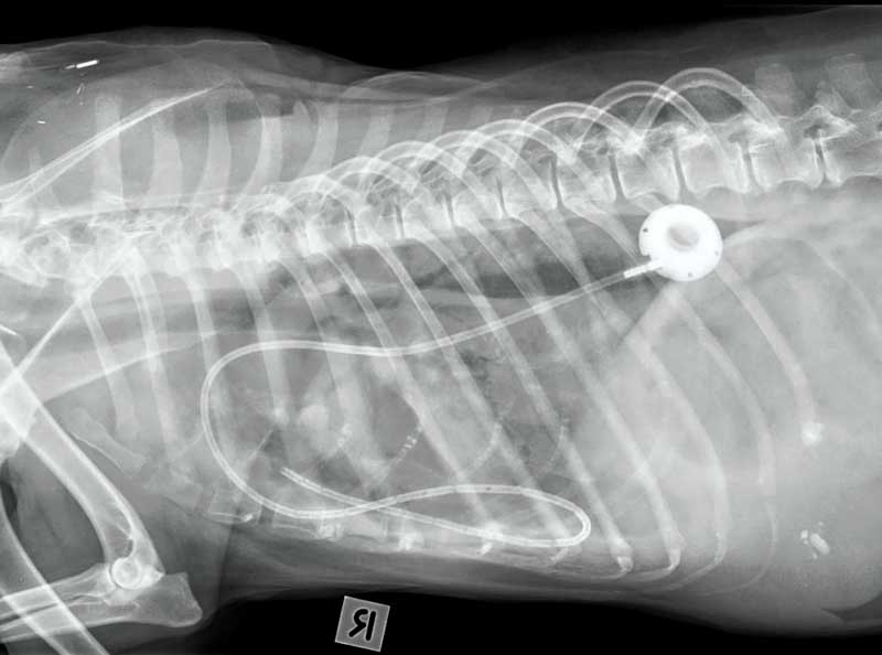
A pleural port kit includes a round-tip fenestrated silicone catheter, which usually is placed in the thoracic cavity, and a titanium port with a silicone septum, placed subcutaneously and connected to the catheter using a boot (Figure 12).
The pleural port is placed surgically and eliminates the need for repeat entry into the pleural space (Figure 13). Access to the cavity is by penetration of the port septum with a specially designed non-corning needle. It allows more drainage for the patient when compared to the conventional TTs. It eliminates inflammation and pain usually associated with repeated thoracocentesis.
Due to the subcutaneous location, the risk of infection is minimised and the risk of pulmonary damage is eliminated.
The pleural port is generally well tolerated and easily located by touch under the skin (Figure 14). It requires minimal maintenance and does not degrade.