21 Aug 2017
In his latest Cytology Corner, Francesco Cian discusses the images from an aspirate from a mass on a digit of a 12-year-old dog.
Francesco Cian
Job Title
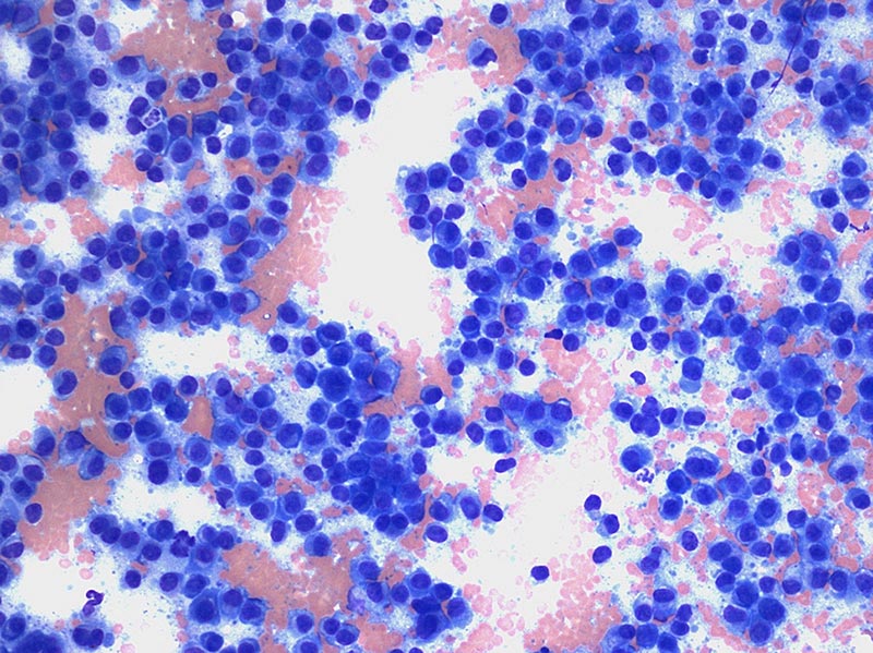
The two images on the right are from an aspirate from a mass on a digit in a 12-year-old Yorkshire terrier (Wright-Giemsa, 10× to 50×). What is your diagnosis?
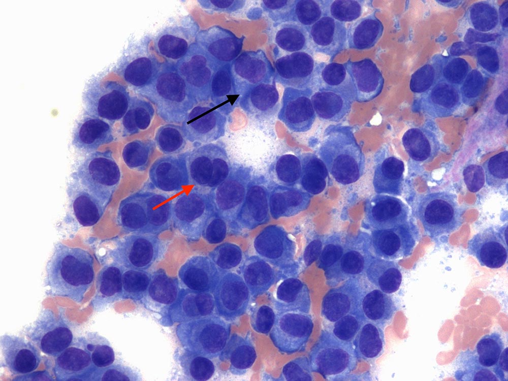
The aspirate is highly cellular, with adequate preservation. On a clear background, with frequent red blood cells, a main population of discrete, round cells is present. These have moderate amounts of deeply blue cytoplasm, with defined borders, often showing a clear perinuclear halo, which represents the Golgi area of the cell (black arrow).
Nuclei are round and paracentral to eccentric, with granular chromatin and poorly visible nucleoli. Anisocytosis (variation of cell size) and anisokaryosis (variation of nuclear size) are mild to moderate.
A few binucleate (red arrow) and trinucleate cells are also apparent, together with rare atypical mitotic figures (not seen in these images). Rare blood-derived neutrophils are also noted.
The submitted aspirate harvested a population of discrete cells and is compatible with a round cell tumour.
The cytological features observed (deeply blue cytoplasm, clear perinuclear halo and eccentric nucleus) are distinctive of a plasma cell tumour and make differentiation from other round cell tumours relatively easy. When this is not possible, histology and immunohistochemistry by using antibodies against multiple myeloma oncogene 1 (MUM1), immunoglobulin light chains, CD79a and CD21 may help to confirm a plasma cell origin.
Plasma cell tumours are relatively common cutaneous neoplasms affecting mainly old dogs and are rare in cats. A breed predisposition exists for terrier breeds, cocker spaniels and standard poodles. Most commonly affected areas include the pinnae, digits, oral cavity and rectum.
Despite the nuclear pleomorphism often observed on cytology and histology (for example, anisocytosis, anisokaryosis, binucleation and multinucleation, and mitotic figures), plasma cell tumours tend to behave in a benign fashion and are usually cured by complete excision; however, a few will recur. Metastases are rare.
Plasma cell tumours may also occur in other sites. When they originate in the bone marrow, they are referred as multiple myeloma; this can be associated with monoclonal gammopathy and hypercalcaemia. Primary extramedullary plasma cell tumours have also been reported in the oral cavity (gingiva), gastrointestinal tract and spleen.