21 May 2024
In the latest of our Diagnostic Dilemmas series, Francesco Cian highlights how erythrocyte morphology assessment via blood smear examination provides important information that can help to establish a differential diagnosis of diseases.
Francesco Cian
Job Title
Image © Sascha / Adobe Stock
A 13-year-old male, neutered flat-coated retriever presented to the referring veterinarian with a history of a few days of lethargy and inappetence.
In-house haematology testing revealed severe anaemia and mild leukocytosis (Table 1).
| Table 1. Haematology results from a dog with severe anaemia | ||
|---|---|---|
| Parameter | Result | Reference interval |
| Haematocrit (L/L) | 0.20 | 0.37 – 0.55 |
| Red blood cell (×10¹²/L) | 2.3 | 5.5 – 8.5 |
| Haemoglobin (g/dL) | 5.7 | 12 – 18 |
| Mean corpuscular volume (fL) | 86 | 60 – 77 |
| Mean corpuscular haemoglobin concentration (g/dL) | 28.1 | 32 – 39 |
| White blood cell (×10⁹/L) | 25.7 | 6 – 15 |
| Platelet count (×10⁹/L) | 280 | 150 – 500 |
A fresh ethylenediaminetetraacetic acid (EDTA) blood sample was sent to an external laboratory for haematology testing and blood smear examination.
Blood smear examination (Figure 1) showed a general decrease of the red blood cell density, confirming the severe anaemia reported by analyser. The anaemia appeared mildly regenerative given the presence of polychromatophils (blue arrow).
Interestingly, numerous crenated red blood cells were present (also known as echinocytes) and variable numbers of schistocytes, which are fragmented red blood cells appearing as irregularly shaped structures, jagged with pointed ends (red arrows).
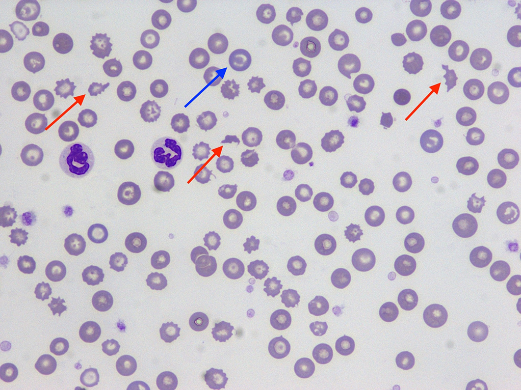
Leukocytes were subjectively increased on the smears, confirming leucocytosis, and were mainly represented by segmented neutrophils (greater than 90%). Platelets were numerous and had unremarkable morphology.
A diagnosis of marked anaemia with signs of regeneration and evidence of schistocytes was confirmed.
The presence of regenerative anaemia suggested haemolysis and/or blood loss as main differentials. Haemolysis due to immune-mediated destruction or oxidative damage were considered less likely given the lack of spherocytes, eccentrocytes and Heinz bodies, respectively.
Haemolysis secondary to red blood cell mechanical damage was considered possible given the presence of schistocytes. These are generally associated with conditions causing mechanical damage to red blood cells due to irregular endothelial lumen, vascular lumen crossed by strands or fibrin and/or turbulent blood flow.
Common differentials in dogs include disseminated intravascular coagulation, glomerular disease, vasculitis, portosystemic shunts and vascular neoplasms (for example, splenic tumours). Echinocytes are very often an artifact that results from excess EDTA, improper smear preparation or prolonged sample storage before blood film preparation.
Abdominal ultrasound revealed the presence of minimal peritoneal effusion and a hyperechoic splenic mass of 4cm in diameter, along with areas of irregular heterogeneous echogenicity.
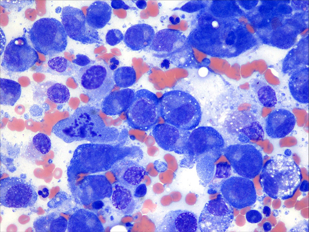
Fine-needle aspiration of the mass showed a main population of individualised atypical cells ranging from round to oval (Figure 2). These cells had variable degree of vacuolation and marked signs of atypia, including anisocytosis (cell size variation), anisokaryosis (nuclear size variation), prominent and multiple nucleoli, and mitotic figures. Few lymphoid cells were also noted alongside blood-derived leukocytes.
The cytological features of the aspirate were supportive of malignant tumour – likely histiocytic sarcoma. Abdominocentesis confirmed the presence of haemoabdomen, but this did not contain neoplastic cells.
To conclude, the reported anaemia was likely due to a combination of blood loss (haemoabdomen secondary to splenic bleeding) and haemolysis (mechanical damage of red blood cells resulting in schistocytosis due to turbulent blood flow caused by the splenic neoplasm). Due to the poor prognosis, the owners elected to have the dog euthanised.
Erythrocyte morphology assessment via blood smear examination provides important information that can help to establish a differential diagnosis of diseases. This information is not provided by automated haematology analysers.