21 Aug 2023
Cytology findings from a stifle swelling in a horse
Francesco Cian DVM, DipECVCP, FRCPath, MRCVS discusses the case of an 11-year-old British warmblood mare examined for a subcutaneous swelling in the cranial stifle region.
Francesco Cian
Job Title
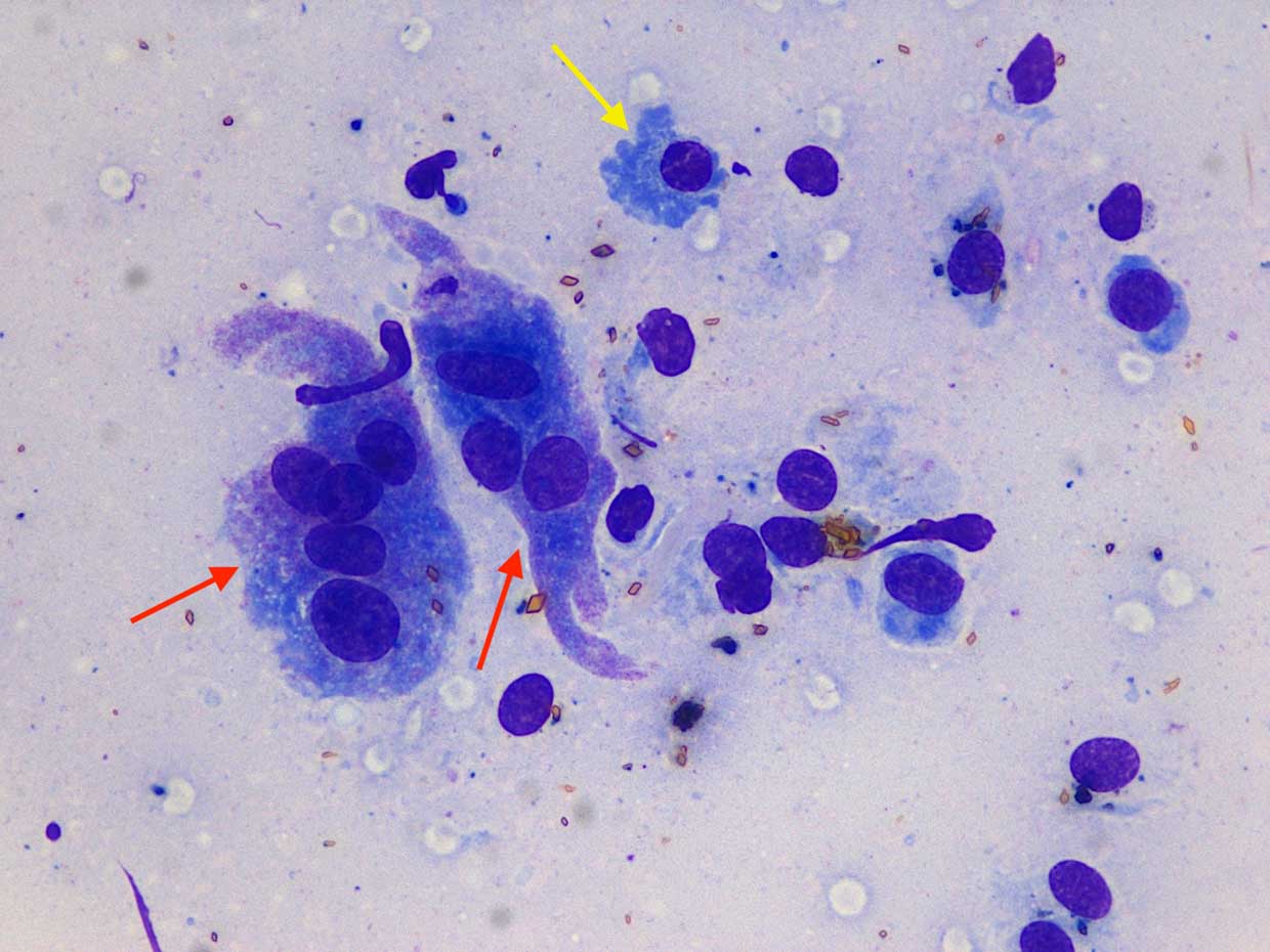
Figure 1. Stifle swelling from a horse, Wright-Giemsa 50×.
An 11-year-old British warmblood mare was examined by the referring veterinarian for a subcutaneous swelling in the cranial stifle region that was first noticed by the owner a few months earlier and had grown in the preceding few weeks.
Clinical examination revealed normal vital parameters and excellent body condition. Ultrasonographic examination confirmed the presence of a hypoechoic mass that was not infiltrating the surrounding tissues. Cytological samples of the mass were obtained by fine-needle aspiration and were submitted to an external laboratory for examination.
The sample had moderate cellularity and excellent preservation. The background was lightly basophilic with small numbers of red blood cells and a few bare nuclei. A mixed population of cells was noted throughout the smears (Figures 1 and 2); these included multinucleated giant cells (red arrows), macrophages often containing haematoidin crystals – yellow rhomboid structures – and products of degradation of red blood cells (pink arrows), spindle cells of mesenchymal origin with mild degree of atypia (blue arrows), and small numbers of other leukocytes, including a few plasma cells (yellow arrow).
Diagnosis
Giant cell tumour of soft parts (GCTSP). These cytological features were considered suggestive of GCTSP.
A granulomatous inflammation with chronic haemorrhage and fibroplasia, possibly secondary to trauma, was also included in the differential list.
Surgical excision was performed and the sample was submitted for histopathological examination, which confirmed the initial cytological diagnosis.
Insight
GCTSP is an uncommon skin tumour of unclear origin, but likely part of the soft tissue sarcoma group; it is reported in different species, in particular cats and horses.
In this species, it most commonly appears as a multinodular, subcutaneous mass in proximity to one of the larger joints of the rear limb, but it has also been described in other sites, such as neck and shoulder.
Microscopically, this tumour is characterised by a mixed population of different cell types, which is unusual for a neoplasm where, generally, a main cell population predominates.
This also represents a challenge of differentiating this neoplasm from a granulomatous-type inflammation and suggests caution in achieving a diagnosis solely based on cytology results. A 2008 review of 21 GCTSP in horses revealed that, in spite of having a malignant histologic appearance, local reoccurrence following surgical removal and metastatic disease were rarely observed.
Take-home message
This case should remind clinicians and pathologists of this unusual soft tissue neoplasm with a predilection for occurrence in the rear limb of horses.