7 Feb 2023
Francesco Cian DVM, DipECVCP, FRCPath, MRCVS presents the case of a referred German shepherd dog with a cervical swelling in his latest Diagnostic Dilemmas.
Francesco Cian
Job Title
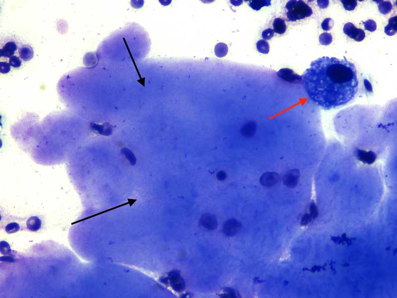
Figure 1a. Neck swelling from a dog (Wright-Giemsa 10×).
A five-year-old male German shepherd dog was seen by the referring veterinarian for a cervical swelling, which had first been noticed by the owner one month before.
On clinical and ultrasound examination, a fluctuant, non-painful, fluid-filled mass was confirmed in the cervical and intermandibular region. Aspiration of the mass under aseptic conditions was performed and the thick mucoid, yellow blood-tinged fluid was collected into a ethylenediaminetetraacetic acid (EDTA) tube and submitted for cytology examination.
The direct smear made from the fluid showed clear background with large numbers of red blood cells and areas of amorphous extracellular lavender material (Figure 1; black arrows). Small numbers of large vacuolated mononuclear cells were present, likely foamy macrophages (red arrow) admixed with rare segmented neutrophils (likely blood derived).
A diagnosis of sialocele was made.
Sialocele, or salivary mucocele, is a collection of saliva that has leaked from a damaged salivary gland or salivary duct, and has accumulated in the tissues.
This is often noted as a fluctuant, painless swelling of the neck or within the oral cavity, depending on the salivary gland involved. The most common types are:
Other less common locations include pharyngeal and zygomatic mucocele.
Clinical signs are often absent unless the mass becomes significantly large and may cause problems such as difficulty eating, or bleeding from trauma.
The cause of salivary mucoceles is rarely identified, although trauma, such as from choke collars, bite wounds or chewing on foreign materials, is generally considered to be the most likely initiating event. As the saliva leaks from the torn salivary gland or duct, it accumulates in the tissue and initiates an intense inflammatory response.
Cytologically, aspirates reveal areas of pinkish amorphous material (mucin/saliva), macrophages (as part of the inflammatory process), neutrophils (blood derived, but sometimes increased as part of the inflammation) and salivary gland epithelial cells. Erythrocytes are usually numerous due to the ongoing haemorrhage that accompanies the salivary gland/duct damage and may show windrowing arrangement due to the high mucin content.
Products of red blood cell degradation (such as haemosiderin or haematoidin crystals) may also be noted as result of (chronic) haemorrhage.
Surgical removal of the affected glands on the side of the mucocele is the normal treatment, and prognosis is usually excellent.
Did you know?
A recent retrospective study conducted by the University of Georgia on more than 170 cases of salivary diseases in dogs showed that almost half of the cases (49.7%) had non-specific sialoadenitis (inflammation).
This was often associated with cavitated areas with accumulation of mucoid secretory material and consistent with a diagnosis of sialocele, likely secondary to duct obstruction by sialoliths. Neoplasia was diagnosed in 20% of the cases and these consisted mainly of malignant epithelial tumours (adenocarcinomas). A surprising number of samples (23%) had no pathologic changes and consisted of normal salivary gland.
This might have been due to incidental aspiration of normal salivary gland tissue in patients with disorders affecting the submandibular lymph nodes as cause for neck swelling. Another possibility for these cases included sialoadenosis, a rare, idiopathic and phenobarbital-responsive disease associated with retching and gulping, and characterised by enlarged salivary glands with no substantial cyto/histopathological changes.