25 May 2015
Isabelle Desmas
Job Title
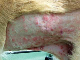
Figure 1. Erythematous, alopecic, ulcerated lesion from T cell epitheliotropic lymphoma.
Canine T cell epitheliotropic lymphoma is a rare disease with a variable clinical presentation. It is characterised by infiltration of cancerous T lymphocytes with a specific tropism for the epidermis and the adnexal structures. Although a preliminary diagnosis can be achieved with cytology, histopathology and immunohistochemistry is required for definitive diagnosis. Solitary lesions can be managed with local treatment (surgery or radiation therapy) if thorough staging has not shown distant spread. However, widespread disease is more common and requires the use of systemic chemotherapy.
The most common chemotherapy treatment described for canine epitheliotropic lymphoma is lomustine and prednisolone. The combination provides a response rate of approximately 80%, with six months median survival. Solitary lesions are rare and long survivors have been described, but canine epitheliotropic lymphoma most commonly presents as a generalised disease poorly responsive to chemotherapy in the long term.
Primary cutaneous lymphoma can be classified as epitheliotropic (epidermal) or non-epitheliotropic (dermal).

Canine cutaneous epitheliotropic lymphoma is more common than the dermal form and is a spontaneous cancer arising from T lymphocytes, which sometimes also involves the mucocutaneous junctions and the oral cavity.
Canine epitheliotropic lymphoma is different from the non-epitheliotropic form of lymphoma (dermal lymphoma) and is characterised by the infiltration of cancerous T lymphocytes with a specific tropism for the epidermis and the adnexal structures (hair follicles, sebaceous and apocrine sweat glands).

Canine epitheliotropic lymphoma is a rare disease that generally arises in middle aged to older dogs. No true breed predisposition has been demonstrated, but English cocker spaniels and boxers seem to be over-represented in the literature3.
As most reports of this disease describe small numbers of dogs and have been written by clinicians from diverse countries, a reference breed population is not available and therefore breed predisposition cannot be accurate. No sex predisposition is reported.
Cutaneous T cell lymphomas are a heterogeneous group of disorders and clinical lesions are quite variable (Figures 1 and 2) – the most common are cutaneous erythema, plaques, scaling and nodules3, but mucocutaneous junctions or oral mucosa can also be affected.
It is mostly a cutaneous disease, but involvement of peripheral lymph nodes and internal organs can be observed in late stages. Different classification systems exist, mainly clinical versus histopathological.
Clinically, the disease can be divided into four categories (Table 1), which are mainly used for descriptive purposes as no link to prognosis has been established in dogs. Three different clinical and histological forms, which originate from the human classification system, have been described – mycosis fungoides, pagetoid reticulosis and Sézary syndrome.

Mycosis fungoides is the most frequent form observed in dogs. It cannot be differentiated from canine pagetoid reticulosis clinically as it is a histopathological diagnosis3.
Sézary syndrome, however, is clinically distinguishable from the others by the presence of neoplastic lymphocytes in the lymph nodes, as well as circulating atypical lymphocytes in the bloodstream.
The clinical presentation may be suggestive of cutaneous lymphoma for experienced clinicians, but the cutaneous lesions are not pathognomonic for the disease. Preliminary diagnosis can be achieved via cytology (direct imprint smear of an ulcerated lesion or fine-needle aspirate of a raised lesion). The final diagnosis is, however, always histologic (punch, incisional or excisional biopsy) because epitheliotropism cannot be identified on cytology.
The lymphoid cancer population is usually monomorphic, but cell size can vary according to histologic subtypes – they are of medium size in the patch/plaque form of mycosis fungoides and of larger size, resembling histiocytes, in the nodular form. In Sézary syndrome, up to 50% of neoplastic lymphocytes are small3.
Immunophenotype can be characterised by immunohistochemistry (or immunocytochemistry) or by flow cytometry. Cancer cells are clusters of differentiation antigen (CD) 3+, a common surface marker of all T lymphocytes. CD antigens are molecules acting as receptors or ligands and present on the cell surface, which provide a target for immunophenotyping of cells. The canine disease is very similar to its human counterpart, but in 80% of the canine cases, cancer cells carry the markers CD4−/CD8+ versus CD4+/CD8− that dominates in 90% of the human patients. In 20% of the cases, the lymphocytes are CD4−/CD8− (natural killer lymphocytes)6,7.
Differential diagnoses include inflammatory lesions such as allergic dermatitis, lupus erythematosus, pemphigus, erythema multiforme and other neoplastic processes, such as cutaneous histiocytoma, Merkel cell tumours or venereal transmissible tumours. Immunohistochemistry for the confirmation of the T immunophenotype can be helpful, but is not specific of a cancerous population.
If the diagnosis is not clear, clonality of the cells can be assessed with a PCR assay7. The PCR for antigen receptor rearrangement should help to distinguish cancer cells (which are monoclonal expansions and therefore share the same DNA) from inflammatory lymphoid cells (which are usually polyclonal). In rare cases a clonal expansion may occur in reactive processes.
A thorough dermatological assessment, as well as complete staging, is strongly advised in cases where localised lesions are observed. Rare cases with solitary lesions have been reported to have long survival, but it is important to ensure there is no widespread disease prior to starting local treatment as sole therapy. In cases where multiple, widespread, cutaneous lesions are observed, dermatological assessment should be sufficient to map the lesions and assess response to systemic treatment, and complete staging is not necessary.
Dermatological assessment should include thorough evaluation of the skin, the interdigital space and the mucocutaneous junctions, as well as the oral mucosa. Palpation of the peripheral lymph nodes and fine-needle aspirate in case of lymphadenopathy should also be performed.
When complete staging is necessary it includes thoracic radiographs and abdominal ultrasound, which are performed to look for internal lymphadenopathy or additional lesions. Fine-needle aspirate of the liver and spleen can also be performed to assess for visceral infiltration, as these organs can look normal on ultrasound, but be diffusely infiltrated by the disease.
The aim of treatment is not a cure, but to achieve remission (disappearance of all lesions) and improve quality of life.

The most effective systemic treatment for canine cutaneous epitheliotropic lymphoma is lomustine chemotherapy. Lomustine is an alkylating agent with a recommended dosage of 60mg/m2 to 90mg/m2, given orally once every three weeks. A total of six doses is generally administered if the disease responds well in the first stage of the protocol. Using this treatment modality, overall response rates of 78% to 83% have been reported in the literature8,9 (Figures 3 and 4); however, the duration of response appears to be generally short (around four months).
Lomustine is commonly given in association with prednisolone, which is beneficial in canine cutaneous epitheliotropic lymphoma for its antipruritic, anti-inflammatory and proapoptotic efficacy. Lomustine is generally well tolerated. Adverse effects associated with its use include myelosuppression, hepatotoxicity, gastrointestinal toxicity and nephrotoxicity. Monitoring with haematology and biochemistry profile is recommended prior to each treatment.
To reduce the likelihood of liver toxicity, it is advised to prescribe a liver protectant (for example, S-Adenosyl methionine and silibinin) for the duration of treatment.
Anecdotal responses to cytotoxic chemotherapy (vincristine, L-asparaginase and cyclophosphamide) and masitinib (tyrosine kinase inhibitor) have been reported in dogs with epitheliotropic lymphoma, but no literature evaluating these treatments on a large scale exists.
Radiation therapy may be beneficial for localised tumours, but complete staging of the disease should be performed before opting for local treatment alone. Information is very limited regarding efficacy of this therapy in dogs with this particular type of tumour. Radiation therapy can also be administered in association with chemotherapy if chemotherapy alone is not sufficient to control the lesions. It may be beneficial when epitheliotropic lymphoma is localised in the oral cavity.
One study established a response rate to radiation of 67%, with a median survival time of 38 months10. In this study, regional lymph node involvement and absence of complete response to the radiation treatment were found to be negative prognostic factors.
Various non-cytotoxic treatments have been reported in the literature, but have not been extensively studied. Linoleic acid (3ml/kg) may be beneficial, but strong evidence is lacking, although six out of eight dogs receiving this treatment improved for up to two years in one study11. Vitamin A analogs have shown success in humans with epitheliotropic lymphoma, but response rates in pets are low and cost, as well as adverse effects, may be an issue12. Human interferon has been anecdotally reported in a dog with epitheliotropic lymphoma with a short-term response of 10 weeks13.
Secondary skin infections should also be treated based on culture and sensitivity results. Patients that are pruritic may benefit from antipruritic topical or systemic therapy (for example, antihistamine medication).
The prognosis is considered poor, with a survival time after diagnosis that ranges from a few months to two years. A median survival time of six months has been reported with lomustine chemotherapy5. Euthanasia is often elected by the owners due to poor quality of life associated with the severity of the skin condition and lethargy. In dogs, no prognostic significance of histologic subtypes has been established. Indolent forms of the disease – generally with localised solitary lesions – have been reported with a better prognosis. In most cases, however, the disease progresses fast and spreads systemically.