16 Jan 2017
Kelly Bowlt Blacklock looks at studies on companion animal wound care, focusing on emerging techniques (part 1/2).
Kelly Bowlt Blacklock
Job Title
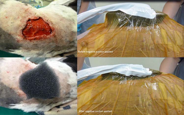
Figure 1. Wound on dorsal lumbar region of a dog. The vacuum-assisted closure system consists of an open-cell foam (400µm to 600µm pore) positioned directly onto the wound bed and covered with airtight dressing. Drainage tubing is connected between the foam and a portable negative pressure suction unit, which contains a single use canister to collect and store any wound fluid removed. When negative suction is applied, the foam visibly compresses. Image: © AHT.
Several outstanding texts have been produced on companion animal wound care that comprehensively examine the basic principles of management1-3.
The mainstay treatment for traumatic wounds usually consists of initial debridement and achievement of a uniform granulation bed using a variety of techniques4. Thereafter, the skin can be encouraged to close by second intention or surgical closure can be attempted1,5.
Research into human and veterinary wound care is constantly evolving. This two-part series will look at some emerging techniques.
Regardless of the treatment used, it is imperative generous, multimodal analgesia should be provided to every patient, discussion of which is outside of the scope of this article.
Importantly, clinicians should also be mindful novel treatments are not always superior treatments – evidence-based medicine should be practised wherever possible.
The most promising development in wound management is negative pressure wound therapy (NPWT), also known as vacuum-assisted closure (VAC). This involves local application of negative pressure to a wound bed via an open cell foam/gauze (Figure 1).
Provision of intermittent or continuous suction is possible via a portable suction unit and evidence exists intermittent NPWT more effectively stimulates angiogenesis and granulation tissue, compared to continuous NPWT6. However, intermittent suction appears to be more painful; therefore, the author uses continuous suction in clinical patients, and generally, a pressure of -125mmHg7,8.
The benefits of NPWT may include reduced oedema and exudate accumulation, increased central wound perfusion9-11, extremely rapid stimulation of a uniform bed of granulation tissue12-16, increased wound fluid pO2, interleukin-8 and vascular endothelial growth factor17 and more rapid wound contraction18. Findings in the literature are inconsistent about the ability of NPWT to reduce wound bacterial contamination19-21.
In the author’s opinion, one of the main advantages of NPWT is the avoidance of the daily anaesthesia/sedation required for traditional debriding techniques, such as wet-to-dry dressings.
The NPWT foam should be changed every 48 to 72 hours because the granulation tissue can grow into the foam if left in place beyond this time, and the less frequent requirement for anaesthesia/sedation allows the patient to increase nutritional intake and ambulate (promoting lymphatic drainage), optimising wound healing.
Dressing strikethrough is eliminated with NPWT because all wound fluid is collected into a single use canister, which is particularly useful in highly exudative wounds.
The most common complications seen with NPWT include mild dermatitis around the wound margin and loss of vacuum; however, these complications are usually readily addressed.
NPWT is widely used in many veterinary hospitals in a variety of cases, including traumatic wounds, decubitus ulcers, degloving injuries, distal extremity wounds13, thoracic or abdominal wound dehiscence, septic peritonitis22, cytotoxic sloughs23, burns24, perineal wounds, osteomyelitis and traumatic wounds with exposed bone (and even shell25), or overexposed orthopaedic implants26.
Interesting case reports/series include:
Several larger studies have been published, looking at the use of NPWT in dogs and cats.
In 45 clinical veterinary patients (dogs), a short time to skin reconstruction was recorded because of the rapid formation of a smooth granulation tissue bed achieved with NPWT7. A total of 96% of the wounds healed after 21 days, despite 11 dogs having previous unsuccessful wound treatment for a mean of 87 days7.
Similar findings of rapid wound healing using NPWT after unsuccessful prolonged management using other techniques have been reported in cats19,34, which have also been reported to suffer fewer complications, less fat tissue necrosis and less sepsis compared with cats treated with a foam dressing. Although a strong effect of NPWT on the progression of infection, fever and sepsis was detected in this population of cats, this difference was not statistically significant34.
NPWT is regularly used in very extensive wounds. In a study by Pitt and Stanley (2014), 50% were greater than 10cm in diameter23.
Finally, a single study has assessed antibiotic concentrations in surgically created wounds treated with NPWT and found the use of NPWT did not statistically impact antibiotic tissue concentrations when compared with conventional non-adherent bandage therapy35. Therefore, any influence of NPWT on sepsis or wound healing in the presence of infection (usually multidrug-resistant in many patients) is not due to increased antibiotic concentration; indeed, the reason for any potential enhanced bacterial clearance with NPWT remains controversial22,21.