9 Jul 2018
Claire Bradley discusses how to spot and treat acidic or alkaline ophthalmic emergencies.
Claire Bradley
Job Title
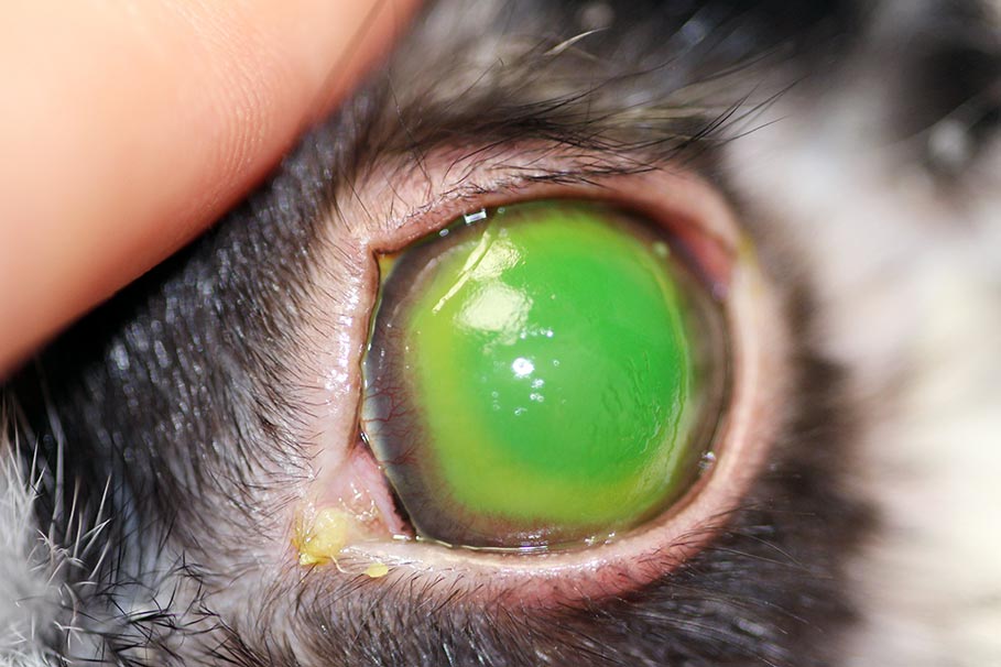
The right eye of a dog 24 hours after contact with an acidic agent. Note the marked hyperaemia of the third eyelid, chemosis, diffuse corneal haze and miotic pupil.
Chemical injury of the canine and feline cornea can lead to sight-threatening consequences. It is important practitioners are aware of the risks associated with such injuries and how best to manage them.
The purpose of this article is to provide recommendations for the emergency management of cases presenting for ocular chemical injury and discuss long-term concerns for these patients.
Although not commonly encountered, chemical injury of the canine and feline cornea represents a true ophthalmic emergency.
It may be sub-classified as acidic or alkaline injury, depending on the causative agent. Examples of acidic agents include sulphuric and hydrochloric acids (found in some drain and toilet cleaners), while examples of alkaline agents include calcium hydroxide (found in some building materials, such as plaster and cement), sodium hydroxide and ammonium hydroxide (found in oven and drain cleaners). It is important to establish whether you are dealing with an acidic or alkaline burn, as they differ in their pathogenesis and overall prognosis.
Owners are frequently unaware their pet has come into contact with an injurious substance – presenting their animal to their primary vet when a painful eye becomes apparent. This highlights the importance of obtaining a thorough clinical history, such as any building work and access to chemical substances.
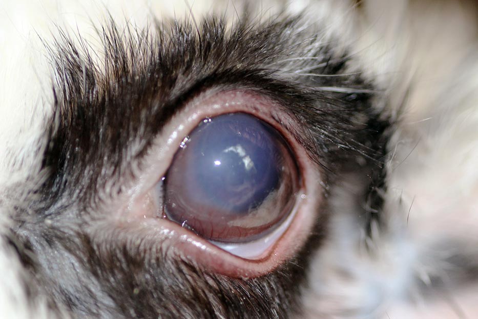
Affected eyes are usually intensely painful in the initial phase, evidenced by marked blepharospasm, eyelid swelling, chemosis and severe conjunctival hyperaemia1. Corneal ulceration is seen with both acidic and alkaline burns. A white ”haze” of the corneal stromal layer is commonly seen in alkali burns. In many cases, the shape of the half-closed palpebral fissure is outlined on the cornea, if the caustic agent was spray delivered.
Where chemical injury is suspected, the use of a pH indicator, such as a urinary test strip, can prove very useful to identify whether the ocular surface is acidic or alkaline. Appreciable differences exist between the pathogenesis of acidic and alkaline burns to the cornea.
Acidic injury: of the two presentations, acidic burns may be considered the lesser of two evils, as these compounds rapidly induce protein coagulation in the corneal epithelium. This results in severe melting of the cornea, but limits access of the acid to the remainder of the eye.
Alkaline injury: alkaline injuries represent a severe threat to vision. These agents readily cross the cornea, and may affect both the anterior chamber and lens2. Alkaline corneal penetration causes stromal keratocyte death. Penetration to deeper tissues may also damage the underlying trabecular meshwork and ciliary body – increasing the risk of glaucoma, uveitis and phthisis bulbi. Alkaline substances react with the corneal stroma, in a process called saponification, which results in swelling of the stromal layer. This manifests clinically as a diffuse white haze to the corneal stroma. Corneal ulceration occurs and may encompass the entire corneal surface. Cases of alkaline injury may develop cataracts, as the noxious agent interferes with lens metabolism.
Both acidic and alkaline injury may result in loss of limbal stem cells. This has severe consequences for the eye, both during the initial healing phase and long term. Stem cell destruction can result in poor corneal epithelial healing, with persistent ulceration and subsequent conjunctivalisation of the cornea3. Symblepharon may also occur. In humans, the degree of limbal stem cell loss is one of a few parameters directly correlated with prognosis4. A classification system for ocular surface injuries does not exist.
Regardless of the causative agent, it is imperative the eye is flushed copiously. Tap water is a suitable diluent and readily available in most instances. Should owners witness the chemical injury taking place, they should be advised to wash the eye with tap water for a minimum of 15 to 30 minutes. Following this, an urgent appointment should be made with a vet.
Once presented to the clinician, a pH indicator strip should advise whether appropriate flushing has taken place (a pH of 7.4 is normal for the cornea). If the pH remains abnormal, continued flushing using designated eye flushes or buffered saline solution should be instituted5, taking care to include the full depth of the corneal fornices. Sterile saline may be used, but has been shown to be less effective at normalising the pH6. Owing to the intense pain, patients will frequently require sedation, or general anaesthesia, to allow adequate lavage of the ocular surface. Particular attention should be paid to removal of any material trapped in the fornices or behind the third eyelid, which may continue to leach alkali/acid.
Animals with chemical corneal burns all experience significant pain in the initial phase of injury. Thus, appropriate analgesia, in the form of opioids and systemic NSAIDs, should be administered. Some patients may benefit from being hospitalised, to enable on-going pain relief.
The use of a bandage contact lens, with or without a temporary tarsorrhaphy, may provide additional analgesia, while the cornea remains ulcerated. Appropriate ocular lubrication should also be provided, as cases of chemical burns have been demonstrated to have subnormal tear production in the initial phases of injury7, presumably due to concurrent damage to the conjunctival goblet cells, and/or the meibomian glands themselves.
Atropine may be used with discretion, to combat the reflex uveitis and iris spasm that may result from corneal ulceration.
Therapy with topical broad-spectrum antibacterials should be instigated, to prevent secondary infections of the cornea. Chloramphenicol is a good choice, although owners should be educated for its off-label use, under the cascade system. In addition, oral doxycycline, at 10mg/kg SID or 5mg/kg BID, may be administered to patients, as it has been shown to help reduce corneal vascularisation, without compromising epithelial wound healing2,8.
Both acidic and alkaline corneal injuries should be treated aggressively with hourly administration of topical serum, with or without ethylenediaminetetraacetic acid. This helps to slow down collagenolysis of the corneal stroma9 – a process that is particularly severe in cases of acidic burns.
Healing of chemical injuries of the cornea may be divided into three phases of recovery4. In the initial phase (day 0 to 7), clinical signs are directly attributable to the severity of the injury. Epithelial regrowth begins, alongside a marked vascular and inflammatory response. During the early repair phase (day 7 to 21), corneal epithelial cells and keratocytes continue to proliferate, and mild injuries may show complete re-epithelialisation. During the late repair phase (more than day 21), mild injuries should be completely healed. More severely affected corneas may undergo conjunctivalisation, stromal scarring or recurrent epithelial erosions.
It is not uncommon to observe initial re-epithelialisation of the ulcerated area of the cornea, which then appears to become denuded again several weeks later. This most likely reflects a lack of ability of new basal cells to re-establish hemi-desmosomal attachments to the basement membrane10. The basement membrane itself may or may not reveal alterations, such as increased thickness. Amniotic membrane transplantation may be beneficial in such cases, to provide an appropriate basement membrane for epithelialisation11-14. However, in severe cases with significant loss of stem cell population, the eye may not be salvageable.
In human medicine, recommendations for treatment have included early, intensive administration of topical corticosteroids to injured eyes2-4. Cases with non-healing epithelial defects are tapered off topical steroids within the first two weeks; however, in the absence of ulceration, this therapy is frequently continued for several weeks. Such treatment, especially in our patients, is not without risk of progression of corneal ulceration, and extreme caution should be taken if treating in this manner. Prior discussion with a specialist ophthalmologist is highly recommended.
It has been shown in animal studies that ascorbate (an essential cofactor for wound healing) is acutely decreased in aqueous concentration, following alkali injury15. Thus, some human physicians now prescribe both topical and systemic ascorbic acid (vitamin C) to affected patients. At present, its benefit in a small animal setting remains unknown.
In human eyes, which are severely and irreversibly damaged by chemical injury, keratoprosthesis implantation, corneal transplantation, and stem cell-based replacement therapy may be considered4. At present, none of these procedures are routinely available in our veterinary species; however, the use of canine keratoprostheses are in their infant stages, for treating corneal diseases, such as endothelial dystrophy, chronic superficial keratitis and keratoconjunctivitis sicca16,17.
To date, one reported case exists of keratoprosthesis placement in a dog with chronic corneal fibrosis following chemical injury17. This patient required further additional treatment following overgrowth of granulation tissue over the optic of the keratoprosthesis. However, follow up at 12 months confirmed a visual eye. What appears radical may well represent a suitable alternative in the future to animals blinded by chemical corneal injuries.
Chemical injury in companion animals poses a potentially devastating threat to vision. Early intervention is of paramount importance and specialist advice should be sought. Patients presenting with an acutely painful eye, with significant corneal ulceration, blepharospasm, eyelid swelling, chemosis, conjunctival hyperaemia, uveitis and a hazy stroma, should alert clinicians to the possibility of chemical corneal injury.
As such, obtaining a full and pertinent history is invaluable, and will help in establishing whether an acidic or alkaline injury has occurred. With prompt veterinary intervention, prognosis for retaining vision may be acceptable in less severely affected cases. Damage to the cornea from an alkaline agent carries a much poorer prognosis and those with significant corneal injury should be referred to a specialist ophthalmologist, if vision is to preserved.
The author wishes to thank Rachael Grundon for reviewing this article prior to publication.