5 Aug 2019
Francesco Cian’s latest Haematology Hub column reviews the case of a dog that presented with loss of appetite, exercise intolerance and a history of persistent anaemia.
Francesco Cian
Job Title
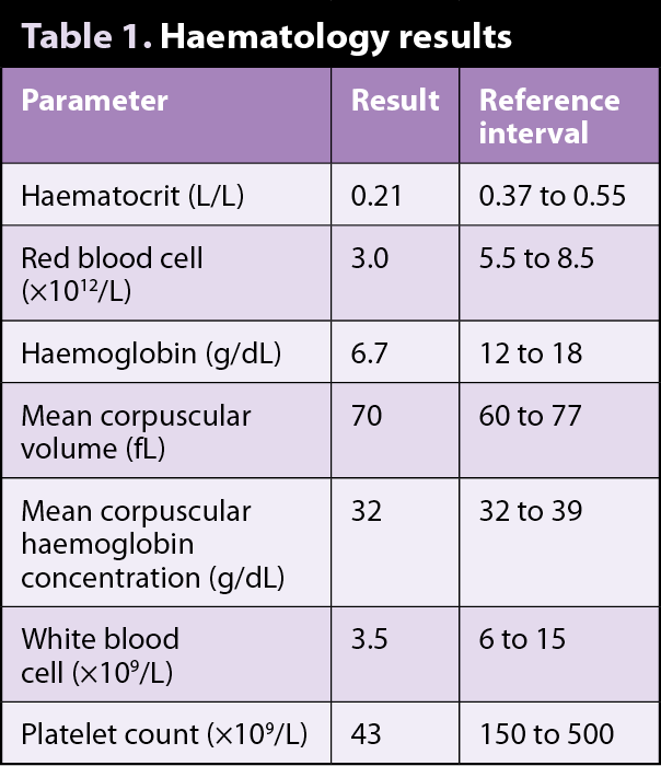
Figure 1. Blood smear (Diff-Quik 50×).
The blood smear (Diff-Quik 50×; Figure 1) and data (Table 1) are from an ethylenediaminetetraacetic acid blood sample of an adult male dog seen by a referring veterinarian for loss of appetite, exercise intolerance, pale mucous membranes and a history of persistent anaemia.
Based on the data and blood smear image provided, try to answer the following questions.
The haematology data shows a marked normocytic normochromic – and likely non-regenerative – anaemia, leukopenia and thrombocytopenia. These results have been confirmed by blood smear examination as red blood cell density appears significantly decreased and no polychromasia is noted.
In most fields, neither white blood cells nor platelets are seen. The decrease in all cell lines in the peripheral blood is referred to as pancytopenia and suggests decreased haematopoiesis due to a primary or secondary bone marrow disease.
When peripheral pancytopenia is present and persistent, and no obvious causes for it are identified, bone marrow aspiration and core biopsy are often strongly recommended.
In most cases, these are able to provide an explanation for the pancytopenia or, at least, reduce the list of differential diagnoses.
The dog was cryptorchid. Ultrasound examination identified a 5cm by 4cm mass in the middle ventral aspect of the caudal abdomen. Fine needle aspiration was performed and a diagnosis of Sertoli cell tumour was made.
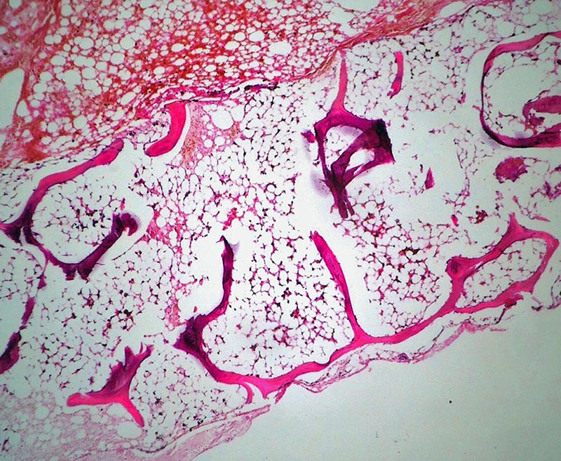
Bone marrow aspiration and core biopsy were collected, and both showed areas with marked decrease of haematopoietic tissue, often replaced by adipose tissue (Figure 2). These findings were considered supportive of bone marrow aplasia due to oestrogen toxicity.
Dogs are susceptible to the myelotoxic effects of oestrogens, when these are administered in dogs or produced by testicular/ovarian granulosa tumours. The mechanism is not fully understood, but it often causes an initial bone marrow granulocyte hyperplasia with neutrophilia, followed by either bone marrow recovery or bone marrow aplasia – as seen in this case – often resulting in peripheral pancytopenia.
In cases of oestrogen-producing tumours, surgical removal of the source may be curative in certain cases.
Both testes were removed from this dog and submitted for histopathology, which confirmed the initial diagnosis. The pancytopenia persisted for the following two weeks the dog came back for re-checks. Follow-up was lost after that.
The term “aplastic anaemia” is often improperly used to indicate a bone marrow condition where all haematopoietic cell lines (red blood cells, white blood cells and platelets) are depleted.
The term bone marrow aplasia is considered more appropriate to describe this condition. This may be caused by infections, drugs, toxins, neoplasia, myelodysplasia, necrosis, osteosclerosis, myelofibrosis, or immune-mediated or idiopathic processes.