4 Sept 2017
Francesco Cian asks for your diagnosis when presented with two images from a bronchoalveolar lavage of an adult male dog with a history of chronic cough.
Francesco Cian
Job Title
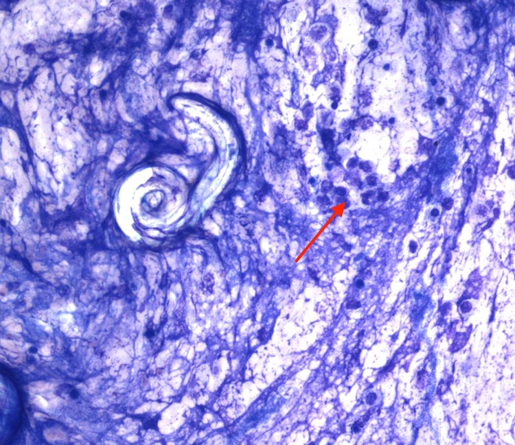
The two images are from a bronchoalveolar lavage (BAL) of an adult male dog with a history of chronic cough (Wright-Giemsa, 10×-50×). What is your diagnosis?
The submitted sample harvested a mixed population of poorly preserved nucleated cells, mostly macrophages and degenerate neutrophils (red arrow) on a basophilic background containing large amounts of amorphous material, cellular debris and extracellular bacteria. Variable numbers of larvae with coiled tails are present, often forming small groups (black arrows).
The cytological features of this BAL are supportive of a lungworm infection. Several types of lungworms affect dogs in the UK, the most common being Angiostrongylus vasorum and Crenosoma vulpis.
Distinction between the two is possible based on morphology, since Angiostrongylus vasorum measures 310μm to 400μm and has a short, kinked tail, whereas Crenosoma vulpis is much shorter (200μm) and has a smaller, pointed, smooth tail. In this specific case, Angiostrongylus vasorum infection was further confirmed on serology.
Angiostrongylus vasorum (also known as French heartworm) is a nematode found in Europe and parts of North America; and is endemic throughout most of the UK.
Adults are commonly observed in the right side of the heart and pulmonary arteries of dogs. Eggs hatch quickly and first-stage larvae migrate through the capillary beds into the bronchioles, bronchi and trachea, causing variable respiratory signs and, in severe cases, bleeding.
A neutrophilic inflammation is commonly observed in BAL samples from infected dogs, with or without concurrent increase in eosinophils. Peripheral eosinophilia and hypercalcemia, the latter secondary to granulomatous inflammation and likely due to the dysregulation of calcitriol production by activated macrophages, are occasionally seen.
Diagnosis depends on the identification of first-stage larvae in the respiratory tract fluid via BAL or tracheal wash.
The Baermann test is typically used to detect larvae in faecal samples. This procedure is considered the gold standard for diagnosis of Angiostrongylus vasorum infection; however, multiple faecal samples should be submitted for analysis in order to avoid false-negative results as excretion of the larvae is intermittent.
Serology testing from serum or plasma is also possible, and has a high sensitivity and specificity, when compared with the gold standard Baermann faecal flotation test. The presence of the antigen in the canine’s blood indicates the animal is actively infected. PCR testing is also available.