23 Nov 2015
John Beel
Job Title
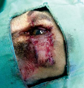
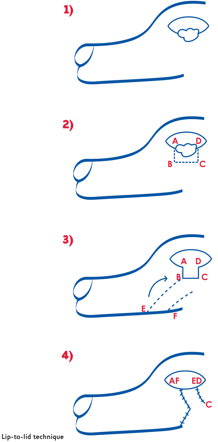
Figure 6. The lip-to-lid technique.
A recently rehomed kitten presented with multiple ocular abnormalities, most notably bilateral upper eyelid agenesis. The upper eyelid defects were corrected in a two-part procedure using a combination of Mustardé technique and lip-to-lid procedure. There was a good cosmetic and functional outcome.

Although not a novel procedure, this case represents a fairly marked congenital defect not commonly seen in practice.
It emphasises the possibilities to correct such significant defects and the possibilities of other further surgical management, including defects created by large neoplasia removal, which is a common presenting problem in first and second opinion practice.
Please note these surgeries should only be undertaken by someone suitably qualified or with considerable experience in eyelid surgery.
The patient was a two-month to three-month-old, male, domestic shorthair cat that had recently been found by its owners as a stray. The owners had noted obvious eye problems and took the cat to their vet.
The referring vet performed a general clinical examination and found the kitten to be in fairly good condition in other respects. It was diagnosed with agenesis of the upper eyelid and initially managed with artificial tears (administered topically three times a day).
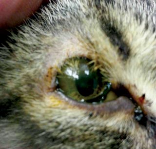
On referral to the author, the patient presented with obvious blepharospasm and mild bilateral mucopurulent ocular discharge (Figure 1). Complete ophthalmic examination revealed 90 per cent to 95 per cent upper eyelid coloboma, resulting in severe trichiasis (Figure 2).
Both eyes were fully sighted and had normal reflexes (menace and pupillary light reflex). The conjunctiva bilaterally was inflamed and showed signs of bacterial infection. The corneas showed significant keratitis, likely from exposure and desiccation (although concurrent viral issues could not be ruled out). Surprisingly, although scarred and having areas of neovascularisation, there was no current active ulceration. The anterior segment had multiple bilateral persistent pupillary membranes. The lenses were present and otherwise normal, as was the posterior segment. The Schirmer tear test was 0mm bilaterally.
A diagnosis of multiple ocular defects was made, with the most significant of these being the coloboma and likely congenital keratoconjunctivitis sicca (KCS).
Options were discussed in detail with the owner, including reconstructive surgical procedures, enucleation and euthanasia.
There were financial constraints. The owners did not have the funds to afford specialist referral, as well as welfare issues with allowing this little guy to continue in such significant discomfort. It was decided we would offer a surgical solution at a much-reduced cost to help the owners and the patient.
After a search of the literature and reconstructive techniques available, we decided to progress with a two-stage procedure.
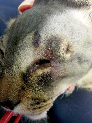
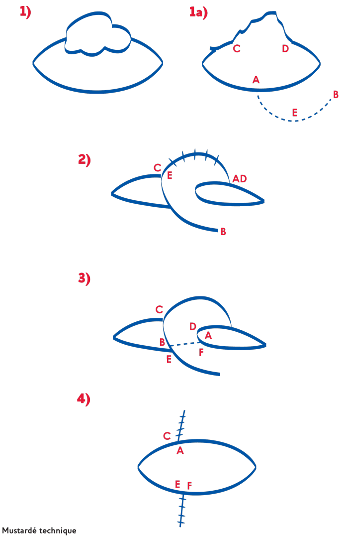
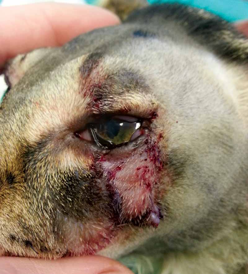
In a second procedure two weeks later, the Mustardé flap was sectioned and the upper eyelid reformed by careful apposition of lid margins into the correct position. The resulting lower lid defect created by this borrowing of the lower lid was then corrected by a single pedicle flap created from the upper lip (a lip-to-lid procedure) and carefully placed to fill the defect (Figures 5, 6 and 7).
The patient handled both operations well and tolerated the temporary blindness while the eyes were closed, and no concerns were raised by its owners.

A small area (approximately 3mm to 4mm) of skin necrosed on the lip graft, but this was not on the lid margin and did not result in any defect of the margin. During the healing period, there was some limited motility of the eyelids, although small superficial ulcerations developed in the centre of the cornea, again assumed to be as a result of exposure and desiccation. These were managed in a routine manner using long-acting topical tear replacers or lubricants and antibiotics.
Over the following few weeks these ulcerations healed and there was progressive functional closure of the eyelids. The Schirmer tear test also improved, assumed to be as a result of better tear retention, reduced inflammation and infection. The tear function did not completely resolve and the patient remains on medical management of this (Figure 8).
The keratitis has almost completely resolved leaving only residual scarring. Comfort and quality of life is good, and the cat’s activity level and habits at 18 months follow-up are those of a normal cat.
Eyelid agenesis or coloboma is a rare anomaly, although more frequently found in cats than dogs1. It is a congenital lesion, often thought to be inherited, and usually bilateral. It is often associated with other ocular defects such as persistent pupillary membrane, lacrimal gland aplasia, cataracts and retinal dysplasias. Eyelid colobomas have been described in every breed, but are more prevalent in Persians2, Burmese3 and domestic shorthaired4.

Treatment options often depend on the severity of the defects and the resulting irritation from exposure and trichiasis. In very minor abnormalities, such as defects involving only the formation of meibomian glands in the eyelid margins, no treatment may be needed and in minor defects one may be able to medically manage the condition with the application of topical lubricants alone.
In larger defects, several surgical procedures have been described with various degrees of success and difficulty. I have mentioned a few of these here for comparison to our chosen technique:
However, even with modifications to include conjunctiva within the flap, this technique does not bring eyelid margin into the defect and does risk trichiasis developing as a result of hair from the flap irritating the cornea.
l Use of various sliding skin and eyelid grafts, for example, z-plasty skin flaps, semi-circular skin flaps and bucket handle technique (Figure 10), can be used, but all have the same problem of a lack of eyelid margin.
We elected to use the combination technique because of the potential to preserve a mucocutaneous junction to preserve the eyelid margin and thus avoid trichiasis.
