15 Feb 2016
Chris Miller
Job Title
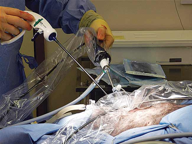
Figure 2. A two-port approach to the abdomen.
Laparoscopy in dogs is becoming an increasingly common technique for procedures such as ovariectomies and biopsies. It allows minimally invasive surgery with enhanced and magnified visualisation of the surgical field (Figure 1).

It is also associated with faster recovery, a reduced risk of postoperative wound complications and significantly less pain than open surgery1,2,3. This article discusses the approach and management of dogs undergoing laparoscopic surgery from an anaesthesia point of view.
To provide suitable visualisation and manipulation of organs the abdomen must first be inflated. Insufflation of the abdomen can either be performed using a blind or open approach. The trochar or Veress needle is inserted and gas insufflated to a pressure of 10mmHg to 12mmHg. The most common gas used is carbon dioxide (CO2) because it is safe to be used with electrocautery and has a low risk of embolism.
Once the abdomen is inflated, more ports can be inserted to allow passage for the laparoscope and instruments (Figures 2 and 3).
The main difference between laparoscopy and laparotomy for abdominal surgery is a raised intra-abdominal pressure (IAP).
Raising IAP using CO2 impacts on the respiratory system mechanically and chemically. The increased pressure will push the diaphragm cranially, producing a splinting of the diaphragm and reducing the functional residual capacity of the lungs.
As a consequence, at normal airway pressures more alveoli will be in a collapsed state, reducing the surface area for gaseous exchange and resulting in less uptake of oxygen from the lungs and reducing the partial pressure of oxygen in the blood.
Areas of atelectasis will still be perfused, leading to a venous admixture further reducing arterial oxygenation. This will impact on the oxygen delivery to the rest of the body and can cause hypoxia and tissue damage.
When CO2 is insufflated into the abdomen some of it is absorbed into the systemic circulation, increasing the partial pressure of CO2 in the blood. The normal physiological response to hypercapnia is to increase the respiratory rate to expire excess CO2. This response in a conscious, healthy animal would be sufficient to manage the increased CO2 concentration in the blood; however, in an anaesthetised animal the response is decreased.
Raising IAP will also have an impact on the cardiovascular system, compressing the caudal vena cava and reducing the venous return to the heart and cardiac output. This reduction in cardiac output may decrease peripheral tissue perfusion, resulting in hypoxia.
The body will respond by increasing the heart rate and systemic vascular resistance to maintain tissue perfusion. However, an anaesthetised animal will have a reduced capacity to mount an appropriate response.
A raised IAP can also reduce perfusion of the organs by compressing abdominal vessels. It has been shown the IAP required for suitable surgical visualisation and working space can maintain normal physiological response cardiac output despite a decrease in venous return.
The most critical period in regard to cardiopulmonary change is at insufflation and desufflation. The sudden difference in IAP has the greatest impact. Interestingly, after long periods of raised IAP, the most dramatic changes are seen at desufflation. A sudden fall in IAP results in increased cardiac output and greater pulmonary perfusion.
However, a greater ventilation/perfusion mismatch causes a decrease in the partial pressure of oxygen in arterial blood (PaO2).
To address the complications of raised IAP you need to detect these changes. Pulse oximetry is vital to assess oxygenation of the blood and provides important information regarding compensatory changes in heart rate as a result of differences in venous return and cardiac output.
Capnography gives an insight into the effects of a raised IAP. Monitoring respiratory rate, pattern and end tidal carbon dioxide (ETCO2) will offer a lot of information regarding compromises to ventilation. A raised ETCO2 would indicate either reduced alveolar ventilation or increased absorption of CO2 from the inflated abdomen.
Normotension should be ensured with a mean arterial pressure >60mHg and systolic >90mmHg. Tachycardia with a low blood pressure would suggest a large reduction in venous return, which is likely to occur in a hypovolaemic patient. Steps to take would include administering boluses of 10ml/kg Hartmann’s solution or the surgeon working at a lower IAP. Arterial blood gas analysis will give a definitive answer of the degree of hypercapnia and acidosis.
To ensure normocapnia, intermittent positive pressure ventilation should be provided, manually or by using an automatic ventilator.
If ventilating manually it is important to understand there will be decreased compliance of the lungs (compliance is the ease with which the lungs can be expanded). Monitoring the extent of chest excursions is vital, albeit a relatively crude method, to prevent generating excessive airway pressures.
The use of a ventilator allows much finer control. Tidal volumes and respiratory rates can be controlled to maintain ventilation. Studies have shown to maintain normal partial pressure of carbon dioxide in arterial blood (PaCO2), minute ventilation needs to be increased and ETCO2 should be maintained at 35mmHg to 45mmHg.
Oxygenation saturation is not dramatically affected by a raised IAP and should not be a major concern if the animal is maintained on 100% oxygen and anaesthetic gas. Peripheral capillary oxygen saturation should be maintained above 93%.
No studies to date have investigated the effects of raised IAP in spontaneously breathing dogs.
Several studies have demonstrated cardiovascular and pulmonary changes during laparoscopic procedures using CO2 as the insufflation gas, but all animals in these studies had been artificially ventilated. These changes tend to be mild and within normal physiological parameters when IAPs less than 15mmHg are used.
In one study, minute ventilation was fixed and PaCO2 did increase as high as 55mmHg4. However, the abdomen was insufflated for three hours and PaCO2 levels did decrease to normal limits 30 minutes after desufflation when minute ventilation was not changed. This suggests, even with fixed minute ventilation, compensation can be made for the changes in PaCO2.
Another study altered the minute ventilation to maintain ETCO2. Again, the abdomen was insufflated for three hours and it was necessary to increase minute ventilation to maintain ETCO2 between 40mmHg to 42mmHg5. However, this increase was within normal physiological limits and not required for the procedure’s whole duration.
If ETCO2 measurements can be made and the abdomen is insufflated at pressures around 10mmHg, a healthy animal should be able to spontaneously breathe and compensate for the increased PaCO2.
Slow insufflation and desufflation are very important in preventing rapid changes in abdominal pressure. Maintaining the animal on 100% inspired oxygen for as long as possible after desufflation will help to minimise the decrease in PaO2 at this stage.
Moderate pain can be associated with insufflation of the abdomen, but there is less postoperative pain using a laparoscopic technique.
An acepromazine/methadone premedication will provide suitable anxiolysis, sedation and analgesia for the duration of the surgery and into the postoperative period. For a healthy dog, appropriate doses would be 0.02mg/kg acepromazine and 0.3mg/kg methadone IM 30 to 40 minutes before induction.
Lidocaine can be infiltrated into the skin and muscles overlying the portal sites at least five minutes before the ports are inserted into the abdomen. Lidocaine patches can be applied over the sites postoperatively.
An NSAID will contribute to perioperative analgesia.
Coagulation times should be assessed before surgery. Acepromazine should be avoided as it may prolong clotting times and increase the risk of haemorrhage.
An alpha-2 agonist, in combination with an opioid, would be an ideal premed. The author considers using dexmedetomidine as opposed to medetomidine as there is less to be metabolised by the liver. In severe cases of liver disease it may be necessary to omit the alpha-2 agonist and rely on an opioid alone.
Appropriate doses depend on the dog, but could be 0.005mg/kg to 0.010mg/kg dexmedetomidine and 0.3mg/kg methadone IM or IV 15 to 20 minutes before induction. All induction agents are metabolised by the liver, but propofol may be the best choice as it is also metabolised by extrahepatic tissues.
Local anaesthetics can be used as described before.
NSAIDs should be avoided and postoperative analgesia should be continued with opioids. There may be a prolonged duration of action of the opioids due to decreased metabolism.
The author would like to thank Matt Gurney and John Williams for reviewing this article and providing the images.