1 Jul 2019
Chris Palgrave and Annedine Conradie present the case of a young cocker spaniel with a malformation on the omphalomesenteric duct.

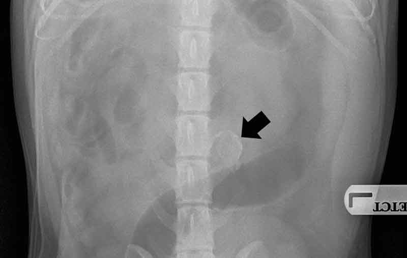
Figure 1a. Left lateral radiograph. An oval, mineralised opacity is present just left of the midline in the caudal abdomen.
A two-year-old male, neutered cocker spaniel presented with a 24-hour history of vomiting. He was uncomfortable on abdominal palpation, and radiography revealed a mineral opacity in his mid-caudal abdomen (Figure 1).
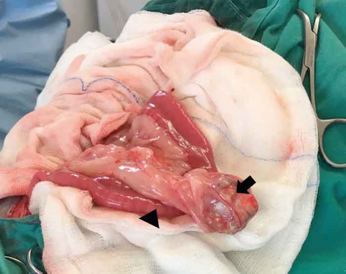
During exploratory laparotomy, a well-demarcated, ovoid, 5cm × 2.5cm × 2cm cystic mass was identified in the mesentery, immediately adjacent to the mid-jejunum (Figure 2). No communication was apparent between the small intestine and the cystic mass.
An enterectomy was performed to remove a 1cm to 2cm section of the jejunum and the adjacent mass.
The dog recovered well, and at follow-up (two months postoperatively) it was clinically normal. The mass was fixed in 10% neutral-buffered formalin and submitted for routine histopathological examination.
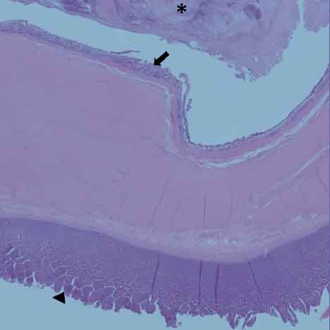
Histopathology revealed a cystic structure lined by well-differentiated, goblet, cell-rich, glandular mucosa forming branching crypts, which resembled colonic mucosa.
The lumen was filled with partially mineralised material, including mucus, sloughed epithelial cells, degenerate neutrophils and pyknotic debris (Figure 3).
The cystic lesion was consistent with a congenital abnormality of the vitelline (omphalomesenteric) duct, which attaches the yolk sac to the developing intestine in the embryo. The duct normally involutes in utero to become a ligamentous cord.
Persistent Meckel’s diverticulum is the most common abnormality of the vitelline duct, often seen in humans, pigs and horses, where it forms a blind-ending sac, which is continuous with the lumen of the small intestine. It is rarely identified in other species.
In humans, a very rare form exists, in which the mid-portion of the duct fails to involute, resulting in a cystic structure that does not connect with the intestinal lumen. Similar lesions have also been reported in a dog, cat and rat.
These rare congenital vitelline duct cysts are typically lined by well-differentiated intestinal mucosa, which may have short villus-type projections, but they predominantly resemble colonic mucosa.
In general, congenital abnormalities of the vitelline duct are considered to be incidental findings. However, in this case, the presence
of granulocytes within the lamina propria and partially mineralised necrotic luminal contents indicates the cyst had become inflamed and/or infected.