4 Sept 2017
Sophie Mahendran looks at diarrhoea in calves and rehydrating those with the condition by administering fluids via the IV and oral routes.
Sophie Mahendran
Job Title
Calf diarrhoea is one of the most prevalent and economically important diseases in these animals worldwide, with up to 20% mortality occurring in some outbreaks of disease (Uetake, 2013).
It’s a multifactorial disease caused by viruses (such as rotavirus, coronavirus and bovine norovirus), bacteria (such as Escherichia coli and Salmonella) and protozoa (such as Cryptosporidium). As always, the success of colostral passive transfer plays a major role in the prevention of calf diarrhoea early on in life, with further management practices and hygiene influencing the occurrence of diarrhoea later.
Physiologically, two types of diarrhoea exist: hypersecretory and malabsorptive. Hypersecretory diarrhoea commonly occurs with E coli infections, which cause dysfunction of cell metabolism or atrophy of the villi, leading to net secretion of chloride, sodium and water into the intestinal lumen. This overwhelms the absorptive capacity of the large intestine and results in increased fluid content of the faeces (Smith, 2009).
Malabsorptive diarrhoea is caused by villi atrophy, resulting in malabsorption of electrolytes and water, which causes an osmotic diarrhoea. The intestinal loss of electrolytes leads to a hypo-osmotic extracellular fluid, which causes free water to move from the extracellular fluid to the intracellular fluid space.
Whatever the causative agent of calf diarrhoea, the end result is generally dehydration and metabolic acidosis from either electrolyte imbalance or D-lactate build up. Traditionally, diarrhoea was thought to result in acidosis from loss of fluid and bicarbonate out in the faeces, but, more recently, the detection of an increase in the anion gap during episodes of diarrhoea led to the discovery of D-lactataemia acidosis (Lorenz, 2009).
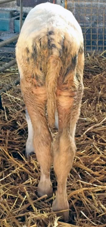
Calves with diarrhoea inevitably have damage to the intestinal enterocytes, allowing undigested carbohydrates to reach the large intestine. This leads to acidification, which favours growth of lactic acid-producing Gram-positive bacteria. It can also be produced following ruminal drinking of milk – although at lower levels. Clinical signs of D-lactataemia include a decreased palpebral reflex, broad-based stance and ataxic movements due to the direct toxic effects on the brain. Signs associated with simple dehydration include a reduced suckle reflex, enophthalmos and an increased skin tent. Generally, perineal staining will also be present, indicating diarrhoea is the causative disease process (Figure 1).
Calf diarrhoea also leads to hyperkalaemia. Initially, this can seem counter-intuitive, as potassium is lost from the body in the diarrhoea. However, potassium is used to buffer the high levels of hydrogen ions found in the blood, along with impairment of sodium/potassium ATPase, which results in increased intracellular sodium and increased extracellular potassium. In addition to this, dehydration leads to aldosterone release from the pituitary, which acts on the kidney to conserve sodium and water, but at the expense of potassium.
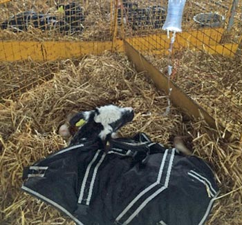
Hyperkalaemia causes impaired neuromuscular excitability and muscular weakness, showing clinically as an inability to stand, and, usually, severe dehydration. These calves will have a prompt response to fluid therapy treatment, unlike calves suffering from D-lactataemia, which takes time to leave the body (Figure 2).
Maintaining calves on milk feeds is strongly encouraged as long as the calf still has a suckle reflex. Using an oesophageal feeder or stomach tube to feed milk is not recommended due to the milk ending up in the rumen, where it may stay if the calf is experiencing ileus, leading to putrefaction and, possibly, bloating. It is worth noting a calf with viral diarrhoea may have reduced lactase activity, which is responsible for hydrolysing the lactose in milk to glucose. This means less glucose – therefore, less energy – is available, but this can be combated by adding lactase to the milk (available in tablet form from chemists) to aid the lactose breakdown.
The addition of bicarbonate directly to milk should also be avoided as this can increase the abomasal pH, which may predispose to increased E coli and Salmonella survival. Also, bicarbonate can react with the acid in the abomasum to produce CO2, thus rendering it unavailable to enter the bloodstream. Acetate and propionate are bicarb precursors added to electrolyte powders to increase the calf’s pH, without affecting the abomasal pH.
The decision to use additional oral rehydration fluids should be made early in the disease course, with there being almost no scenarios when the addition of extra fluids will be detrimental to the health of the calf involved. In the author’s experience, the application of one or two extra oral feeds on the first day diarrhoea is spotted – even if mild – can have a big impact on the calf’s ability to recover quickly.
In addition to this, NSAIDs can be useful to reduce the gastrointestinal cramping and discomfort many of these calves feel, leading to increased voluntary feed intakes. However, care must be taken in the use of NSAIDs in severely dehydrated calves and with repeated treatments of NSAIDs, which can lead to abomasal ulceration and possible renal effects.
Oral rehydration should be used when calves are less than 8% dehydrated, but still have a suckle reflex (Lorenz et al, 2011). If a calf is recumbent, it is likely to be more than 10% dehydrated and will require IV fluid therapy. The main aims of oral rehydration fluids is to promote plasma expansion, correct electrolyte imbalances, provide energy for sodium and water uptake from the small intestine, and act as an alkalising agent to address metabolic acidosis. Four generations of oral rehydration fluids are available, with each having a slightly different electrolyte composition and, therefore, indications for use.
Oral electrolytes can be given as extra feeds, with milk offered at regular feeding times in the morning and afternoon, and with additional electrolytes given at lunch time and the evening. If calves are not willing to suckle milk feeds, these meals can also be replaced by electrolyte feeds, with the use of third or fourth generation solutions providing energy for short-term maintenance.
(Deficit × kg bodyweight) + (maintenance 50ml/kg to 100ml/kg bodyweight/day) = litres fluid calf needs to receive over a 24-hour period.
Can also add 100ml/kg/day to cover ongoing losses from continued diarrhoea.
For example, a 50kg calf that is 10% dehydrated needs: (0.1 × 50kg) + (50ml × 50kg) + 100ml × 50kg = 10L needed over 24 hours.
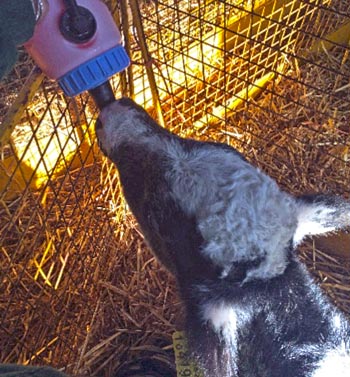
Electrolyte solutions can be given via a teat bottle, if the calf is willing to suckle, or via an oesophageal feeder. Although the latter option is much quicker, the author strongly encourages taking the time to get calves to suckle, with repeated use of a feeding tube causing irritation to the pharyngeal area, along with calves becoming stressed and resenting future handling.
IV fluid therapy is generally required when calves are more than 10% dehydrated, or if they are suffering from metabolic D-lactataemia acidosis. The use of Hartmann’s solution or 0.9% saline is common, but additional potassium should be avoided as the calf will initially be hyperkalaemic due to the hydrogen/potassium ion exchange that occurs. Jugular catheters should be sutured and bandaged into place to prevent the calf rubbing it out, and the calf should be isolated in a small space, ideally with a heat source, along with pre-warming the fluids before administration.
A calculation for fluid requirements is shown in Panel 1, with half of the fluid imbalance being corrected over the first 6 hours and the rest over 24 hours. Additionally, you can give 80ml/kg/hour for up to 30 minutes initially as a bolus (open the drip up).
Amount (mmol) of bicarbonate to give = bodyweight (kg) × base deficit (mmol/L) × 0.5, with 1g of sodium bicarbonate = 12mmol.
For example, a 50kg calf needs 50kg × 10mmol/L × 0.5 = 250mmol of bicarbonate. 250mmol ÷ 12 = 21g of bicarbonate administered over 6 to 12 hours.
If a calf is suffering from metabolic acidosis, IV fluid therapy with bicarbonate-containing fluids is required. You should aim to correct the acidosis over a 6 to 12-hour period, with caution required if measurement of the acid-base balance and base deficit is not possible. Generally, a base deficit of 10mmol/L is a safe figure to use, with rehydration of the calf allowing the kidneys to further correct any remaining acidosis (Panel 2).
Calf diarrhoea is an expensive and time-consuming disease to deal with – both due to the extra care and attention these sick calves require, along with the reduction in growth rates and possible death of those affected.
Prompt administration of oral fluid therapy can be instrumental in reducing losses and speeding up the time to recovery, as well as preventing the more severe secondary consequences of acidosis and hyperkalaemia, which require IV fluid therapy.
Farmers should be strongly encouraged to use the available electrolyte solutions, taking note of the energy levels provided by each type and coming up with strategies to use them effectively on their own farms.