13 Aug 2018
In his article, James Dixon provides a precis of some of the more common causes of lameness in livestock, summarising best advice for treating and preventing them.
James Dixon
Job Title
Figure 1. Infectious lameness in cattle is primarily due to the treponemal infection digital dermatitis.
Lameness is a common problem across cattle and sheep in the UK and is extremely damaging to livestock welfare, production and, ultimately, profitability.
Lameness aetiologies are often split into infectious and non-infectious causes, which often have some common principles when we are considering risk factors and control strategies.
Both types of lameness are prevalent in cattle, especially dairy cattle, whereas it is generally accepted most lameness in sheep has an infectious aetiology. This is likely to be due to their lower bodyweight, and the fact they are generally kept at pasture, rather than on concrete like dairy cows.
Although the manifestations and risk factors are not all identical, many of the causative agents are the same for both cattle and sheep, as are many of the principles behind their control.
Infectious lameness in cattle is primarily due to the treponemal infection digital dermatitis (DD; Figure 1), with the same bacteria also implicated in contagious ovine digital dermatitis (CODD) in sheep.
Fusobacteria necrophorum is known to cause interdigital necrobacillosis (foul in the foot) in cattle, as well as potentially having a contributory role in the scald/footrot complex in sheep.
Treponemes are a form of Spirochaete that is a phylum of bacteria characterised by their spiral appearance and implicated in a wide range of diseases from syphilis to Lyme disease. To date, at least three treponemes have been associated with the characteristic DD and CODD lesions, either alone or in combination.
CODD in sheep is characterised by an erosive/ulcerative lesion, beginning at the coronary band and progressing under the hoof capsule, where it causes severe under-running and, in some cases, complete loss of the hoof horn.
A similar process is recognised in cattle, with the DD lesion at the coronary band and then an infected tract running down below the hoof horn and often progressing to the condition known as toe necrosis. However, the most common presentation of DD in cattle remains a small to medium-sized lesion of the interdigital skin, usually between the heel bulbs.
The different stages of this have been defined using the “M” system of scoring, with lesions developing from a small area of redness/ulceration (M1) to a larger, painful M2 lesion, then a proliferative M3 and finally to a resolving M4 lesion.
Due to the insidious nature of the treponemes and their ability to counteract local host immune defences, a dormant or carrier state, known as M4.1, exists that is thought to play an important part in epidemiology of the disease, with these carrier cows acting as a reservoir of infection for the rest of the herd.
Treatment of both CODD and DD can include a combination of parenteral and topical treatments, with oxytetracycline spray still the most popular, but non-antibiotic treatments, such as Intra Repiderma Spray, are also effective.
The full resolution of these lesions requires prolonged treatment and this poses the greatest challenge to the farmer, with affected animals needing to be caught, restrained and treated daily for up to seven days.
Foul in the foot is an acute condition in cattle caused by F necrophorum, which gains access through damaged or compromised skin and leads to a painful, swollen necrotising lesion. It can affect cattle in all systems, with wet conditions, excess slurry depth and turnout on to spiky grass, such as silage aftermath, all potential risk factors as they traumatise the interdigital skin and allow entry of the bacteria.
Treatment is with prompt antibiotic therapy (simple penicillin is still very effective), combined with NSAIDs to reduce the swelling and associated pain. Resolution is rapid and complete when treated early enough, although some cases can be peracute in onset and require rapid, aggressive therapy.
These conditions are now widely considered as two different manifestations of the same problem, with Dichelobacter nodosus as the causative agent in both cases. D nodosus is a Gram negative, obligate anaerobe with a particular affinity for the epidermal cells of the hoof matrix, where it causes a painful, under-run lesion – with a foul smell and sloughing of the horn capsule a common sequelae.
Where the infection is of the interdigital skin (most common in lambs), the characteristic scald lesion develops with a moist area or erythema between the cleats, often covered with a white pasty plaque. It cannot survive for long in the environment, meaning infected sheep are the primary source of infection and those with chronic/repeat cases pose the greatest risk to their flock mates.
Treatment of scald is usually with topical oxytetracycline spray, which is very effective. Treatment of footrot requires both topical and parenteral antibiotics for a minimum of three days, and it is recommended affected sheep are removed from the flock until treatment has been successful to limit the spread of disease to others in the flock.
The successful control of any of the aforementioned infectious foot complaints relies on correct application of the basic principles of infectious disease control, as would be used, for example, for a BVD problem within one of our cattle herds.
With a number of different agents implicated in these conditions, including three different treponemes and 19 different strains of D nodosus, all livestock farmers should be mindful of the risk of importing further problems, either with bought-in stock or via some of the usual fomites, for example, wellies, equipment or fodder.
With infected animals forming the principal reservoir of infection for footrot, scald, CODD and DD, no control programme can be effective without tackling them. In the case of sheep, infected animals should be separated for treatment and only returned to the main flock when lesions have resolved.
In a dairy herd, where this may not be practical, so-called “blitz therapy” has been shown to be effective by hitting all infected cows at the same time, leading to a rapid decrease in the infectious load within that herd.
This is done by doing an in-parlour DD score of the herd and then shedding out all infected animals for treatment. After an initial treatment in the crush, they can be marked for repeat spraying in the parlour each day.
Repeat offenders with any of these conditions should be culled, as they present a substantial infection risk to the rest of the herd/flock.
Susceptibility to disease is influenced by many factors, such as environment, genetics or nutritional status.
In the case of footrot, resistance can be improved through genetic selection, but also by using the Footvax vaccine at known risk periods, for example, late summer/autumn and spring.
In the case of DD in cattle, it relies much more on environmental management to keep feet as clean and dry as possible to prevent the macerating effect of slurry on the interdigital skin. Strategies here include regular scraping, wider passageways and good cubicle comfort to ensure cows lie down quickly.
Efforts are ongoing to produce a DD vaccine, which may well be an important tool in the control of this disease in the future.
Even with all the aforementioned measures, it is likely some bacteria will remain and this is where footbathing programmes come in, to improve foot hygiene through disinfection and prevent spread within the herd/flock.
Many agents are available for this, with formalin still the most popular and one of the most effective. It is, of course, a very unpleasant substance to work with, and we should take great care to ensure clients will handle it correctly and that the footbath is away from people and well ventilated.
Copper sulphate is an excellent alternative, and, while it is more expensive, usage can be reduced using acidifiers such as Healthy Hooves, which increase the availability of the copper within the solution, meaning less is required to achieve the same result.
A thorough review of footbathing protocols is beyond the scope of this article, but the key principles include correct dimensions, good choice of chemical, accurate dosing and regular refreshing of the fluid.
As mentioned in the introduction, these claw horn lesions are primarily found in cattle due to their increased weight and exposure to concrete, although sheep are occasionally seen with white line abscesses, due to wet conditions or stone damage.
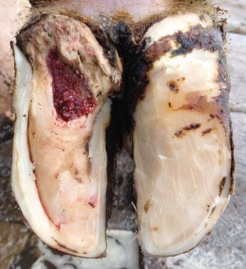
Sole haemorrhage and sole ulcer (Figure 2)are different severities of the same lesion, and are caused by a pressure injury to the corium that is responsible for producing the solar horn. This is most common at the “typical site” in the caudal third of the sole, which corresponds to the point of insertion of the flexor tendons on to the pedal bone.
Due to a growing body of research, we now know a clear association can be seen between low body condition, or rapid loss of condition, and the risk of developing a sole ulcer. This is thought to be due to the shock-absorbing effect of the fatty digital cushion, which is lost in thin cows, meaning the corium is exposed to greater concussive forces.
Sole ulcers should be treated by trimming the foot using the five-step Dutch method, which includes lowering the affected heel and dishing out the typical site as much as possible to alleviate the pressure.
Strong evidence now shows placing a block on the unaffected claw and treating the cow with three days of ketoprofen will have a beneficial effect on the cure rate, especially in early cases.
Prevention of ulcers should focus on cow comfort, standing times and effective feeding and management to prevent loss of body condition after calving.
The white line is the junction between the solar horn, growing from the corium, and laminar horn, growing down from the coronary band, and can be a point of weakness where foreign material is able to gain access if the hoof is placed under undue stress.
When this happens, an abscess is set up within the hoof capsule, and due to the level of inflammation and the lack of room for it to disperse, this is often an acutely painful lesion, with some cows unwilling to bear weight at all. If left untreated, the infection will follow the path of least resistance and often bursts out at the coronary band.
Recognition and treatment are, again, reliant on the five-step method, with the removal of all loose and under-run horn a critical component of this to ensure adequate drainage for the pus and prevent further tracking of the problem within the hoof.
As for sole ulcers, the affected claw should be blocked where possible, and three days of ketoprofen administered.
Control and prevention of WLD also focuses on managing body condition, but also on preventing excessive forces being applied to the feet, and ensuring all cow tracks and floors are well maintained and grooved to prevent slipping and stone damage.
Biotin in the feed has also been shown to help with WLD incidence, but must be fed for at least seven months to see the benefit.
This has been a whistle-stop tour of some of the more common causes of lameness in sheep and cattle, and it is by no means an exhaustive list. The key message to take home is that lameness control must be tackled in a holistic fashion, with all the risk factors considered and tackled where appropriate.
Of crucial importance, in both sheep and cattle, is the early detection and treatment of clinical cases, both to maximise their chance of being cured by the treatment, but also to reduce the infection pressure across the herd/flock.
Other preventive measures can then be focused on risk factors most known to pertain to the kinds of lesions being seen.