11 Mar 2019
Kaz Strycharczyk describes techniques to deal with this common emergency situation, including aftercare and prognosis.
Kaz Strycharczyk
Job Title
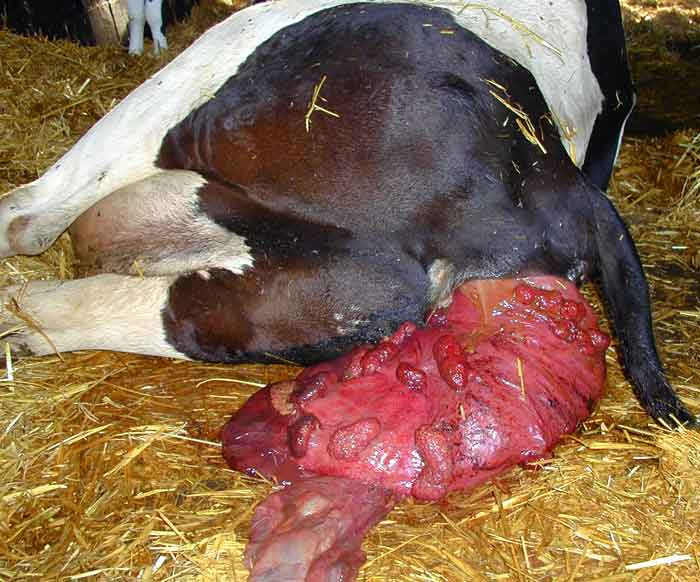
Figure 1. This uterus is relatively clean, although it could have been wrapped in a damp sheet to protect from further soiling or laceration. Image: Julia James / Larkmead Veterinary Group
Uterine prolapse is a common and, if untreated, fatal disease of cattle. This article serves as a refresher in the treatment, control and implications of the condition. Prior to treatment, it is vital to assess if treatment is a suitable option and, if so, which treatment is most appropriate.
In most cases, treatment consists of restraint, replacement of the uterus and adjunctive drug therapy. The specific drugs used vary between practitioner and case, but oxytocin, antibiotics and anti-inflammatories are typically recommended. The finer details of treatment – the need for vulval sutures, or the timing of calcium supplementation – are more controversial. Diligent aftercare, including effective nursing, is also critical. In extreme cases, euthanasia or amputation may be indicated.
A multitude of risk factors for uterine prolapse have been suggested and, of these, dystocia and hypocalcaemia have the greatest evidence base. Prevention measures, therefore, are directed towards these conditions, in addition to sensible and proportionate general husbandry techniques.
The prognosis of uterine prolapses attended rapidly is positive, in terms of both survival and fertility. The condition is also unlikely to relapse – either in the short term or at subsequent calvings. Prompt treatment, therefore, of the uterine prolapse can be cost-effective as well as beneficial for the cow’s welfare.
Uterine prolapse of the cow has a well-earned reputation for eliciting dread in vets. The condition combines an unholy trinity: it is common, requires urgent correction and demands robust upper body strength. Even Alf Wight wrote about the condition in unmistakably bitter tones.
However, a broad veterinary consensus exists in treatment protocols for uterine prolapse in the cow. This article is a step-by-step guide to decision making and correction of this prolapse. The aetiology of uterine prolapse is also discussed with potential measures to reduce incidence.
A uterine prolapse qualifies as a farm animal emergency because of the catastrophic potential sequelae – laceration of the prolapsed uterus, uterine artery rupture and circulatory shock among them. As such, cases should be attended to without delay.
Once on-farm, a brisk – but thorough – assessment of the cow will allow the vet to make the best choice of treatment options.
The following factors should be assessed:
Consideration should also be given to the calf, if present. It is widely accepted most uterine prolapses occur within 24 hours of parturition, and occur more often in cases of dystocia. If treatment of the cow may compromise the health or welfare of the calf, or if the calf is already compromised, appropriate measures should be taken to ensure calf survival.
In cattle that have a poor systemic outlook (for example, in severe shock), severe laceration with protruding viscera, or a low economic value, the costs of intensive treatment will be significant with a low chance of recouping losses. This should be made clear to the owner (Figure 1).
Provided the animal is a viable candidate for treatment (based on the assessment discussed previously), the following protocol may be a suitable template. Similar protocols are discussed by Andrews et al (2008) and Potter (2008).
1. During the telephone call, encourage the farmer to isolate and restrain the cow if feasible. The uterus can be wrapped in a clean, damp sheet to prevent further damage.
2. Once arrived, perform a caudal epidural. Using 5ml of 2% procaine typically has the desired effect.
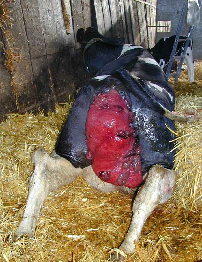
3. If standing, a gate may be used to hold the cow steady. If recumbent or cast, place the cow in the “New Zealand” position with legs behind in the manner of a frog (Figure 2). In this position the vulval lips may be parted and the tail moved out of the way by an assistant if present.
4. Gently lavage the uterus with saline and place on to a clean sheet if recumbent.
5. If readily detachable, the fetal membranes may be removed by gently peeling away at the cotyledons. Otherwise, leave the fetal membranes in place, as traumatic removal is counterproductive. If left in situ these membranes can form a protective layer between the uterus and the environment.
6. Repair full-thickness damage with absorbable sutures.
7. Raise the uterus above the vulva and replace through the vulva. Using ample obstetrical lubricant, gently massage the uterus in. Use palms of hands or fists to avoid perforating a fragile uterus. Replace the body of the uterus followed by the horns.
8. If recumbent, encourage the cow to rise. Fully replace the tips of the horns to ensure full inversion – this is when a wine bottle traditionally comes in useful.
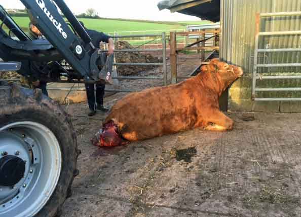
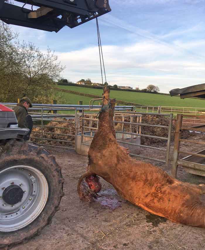
If the uterus is particularly difficult to replace and the cow is recumbent, the hindquarters of the cow may be raised carefully using straw bales or with very careful use of soft leg ropes and a tractor (Figure 3). Ishii et al (2010) describe the use of a tractor in this way with 13 cows with no adverse events reported and a 69% recovery rate (compared to 60.6% across all cows in the study).
If the uterus is bleeding severely, the source should be identified and ligated as soon as possible. Uterine arteries may rupture in dystocia or as a result of trauma after prolapsing the uterus, leading to exsanguination (Divers and Peek, 2007). Again, swift attendance to the case is paramount in improving survival.
Suturing the vulval lips is not universally practised (Noakes, 2009). Proponents believe that even if the sutures fail to prevent a prolapse, they delay and reduce the extent of a re-prolapse, and so reduce the work needed to replace it for a second time. Detractors may believe they are unnecessary or just a “placebo for the farmer” (Roberts, 1982). Alternatively, they believe the sutures cause more straining, or the potential for sutures to tear out during a re-prolapse overrides any potential benefits.
The paucity of trial data means the debate will, no doubt, continue. However, the risk of sutures tearing can be mitigated by using 3mm to 6mm nylon tape with external stents. Where sutures are placed, they can be inserted using the Buhner method – although some vets have modified this technique to incorporate partial exposed horizontal mattress-like sutures into the pattern (Pittman, 2010). Advocates of this latter technique claim it has a reduced stitch failure.
If practising in a resource-poor setting, other suture materials may be used. An Indian study reported the successful use of sterile infusion drip sets as retention sutures (Bhattacharyya et al, 2012); no tearing or recurrence was noted in 44 cases of uterine prolapse. The suture was removed from 48 hours, providing no recurrence took place.
In cases where the uterus is very fragile or has undergone extensive necrosis, an alternative and radical treatment option is amputation.
The following technique has been described:
1. Administer a caudal epidural (as previously mentioned).
2. Place a tourniquet at the vulval lips.
3. Starting 5cm to 10cm from the tourniquet, make a full-thickness dissection of the uterus.
4. Close the wound edges by oversewing.
5. Keeping the tourniquet in place, invert and replace the uterus.
Literature on survival post-amputation is not forthcoming, but the prognosis is regarded as poor to guarded by Weaver et al (2013) who also describe two alternative techniques. Haemorrhage and subsequent shock are serious complications associated with this procedure.
Aftercare should be intensive in these cases, not least because the cow is likely to be periparturient. Both cow and calf should be closely monitored for any signs of deterioration in the immediate post-correction period. Sensible husbandry, including sympathetic nursing and provision of a light, palatable diet, will aid recovery.
As uterine prolapses are associated with dystocia or assisted calvings, make sure the farmer is aware of other possible sequelae, such as retained fetal membranes or necrotic vaginitis. Additionally, the following medications should be considered:
In any case, high risk individuals probably warrant calcium at some stage. The systemic health of the cow is likely to be key in determining whether calcium is given before or after replacement of the uterus; a cow in stage-three milk fever probably requires calcium more urgently than uterine eversion. If in doubt, a blood sample can always be taken for later analysis.
Uterine prolapse follows approximately 0.3% to 0.6% of all calvings (Correa et al, 1992; Plenderleith, 1986), while assisted calvings carry a higher risk (Plenderleith, 1986). As in ewes, the abundance of suggested risk factors suggests a complex aetiology. Factors supported by the relevant literature include:
Other proposed factors include feto-maternal size mismatch, excessive traction in assistance, over-conditioning of animals, high oestrogen content in diet and straining secondary to post-parturient trauma to the reproductive tract.
Studies examining the effect of parity on incidence of uterine prolapse have yielded mixed results. Correa et al (1992) and Gröhn et al (1990) found no significant association, while others have found incidence increased with parity (Markusfeld, 1987). The increased risk of hypocalcaemia in older cattle (DeGaris and Lean, 2008), due to a decreased capacity to mobilise calcium from bone, may explain this potential association.
A lack of clear, correctable risk factors makes preventive measures difficult to draw up. Additionally, unconvincing potential gains may hamper compliance. Therefore, the best prevention measures may be those farmers could already be integrating into the control of other conditions. These would include body condition scoring to avoid over-conditioning, use of a bull with a high easy calving estimated breeding value especially for heifers, appropriate supervision in the periparturient period and, if necessary, prompt, but sympathetic, assistance in a difficult calving.
As hypocalcaemia is associated with prolapses due to its effect on uterine tone, problem farms may benefit from metabolic profiling to ensure cattle are not subclinical deficient. Where hypocalcaemia is identified, a number of preventive measures are available: low dietary cation-anion difference diets, low calcium to phosphorus ratio diets in the final three weeks of pregnancy and oral calcium supplements at calving.
The mortality for uterine prolapses attended quickly is significant. Gardner et al (1990) reported 27.6% mortality within two weeks for American dairy cows treated with a similar protocol as described previously. This paper also reports a significantly improved survival rate if the calf was born alive, the cow was primiparous, or the cow was not in stage-three milk fever at the time of treatment. The prognosis for cows treated by amputation is likely to be significantly poorer than average.
Jubb et al (1990) found a similar proportion of cases survived (73.5%, or 26.5% mortality) in Australian dairy cattle. Both this study and Oakley (1992) found no incidence of relapse. They also followed cows through to the subsequent pregnancy and both found acceptable rates of conception, although the former found uterine prolapse cases took 10 days longer to conceive than controls.
In American beef cattle, however, prolapse has been found to reduce subsequent pregnancy rates (Patterson et al, 1981), although this study amalgamated vaginal and uterine prolapses.
Uterine prolapse in cows is an emergency that warrants urgent treatment on welfare grounds. Additionally, when viable, treatment typically returns the cow to a productive and fertile life during which the prolapse is unlikely to recur.
The basic protocol involves assessment, restraint, replacement and adjunctive therapies. Vulval retention sutures remain the subject of debate. Risk factors supported by experimental data are dystocia and hypocalcaemia. Therefore, on farms experiencing high incidence of uterine prolapse, stock management should be investigated for any practice, or lack thereof, that may contribute to either of these.
The most appropriate preventive measures may well be those with wide-ranging benefits and so should be encouraged as part of any stockman’s management plan.