20 Nov 2017
David Rendle outlines the causes, development, symptoms and treatment options for this neurological condition in horses (includes video content).
David Rendle
Job Title
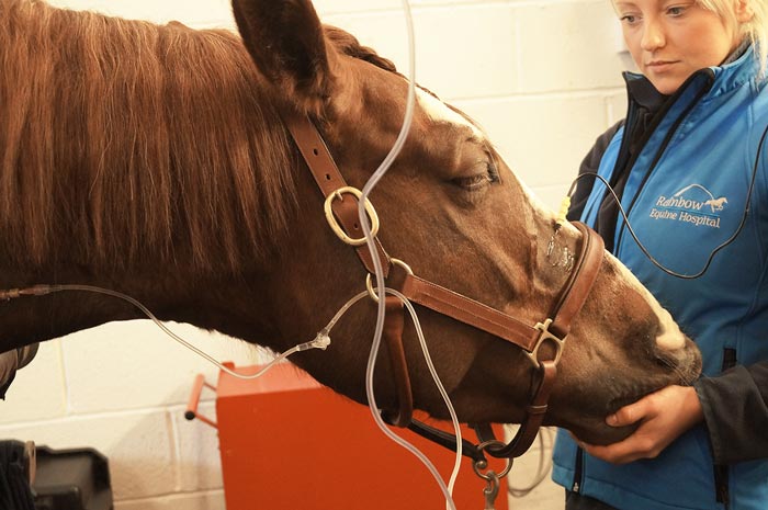
Figure 1. CT provides the best means of imaging the head to eliminate potential causes of headshaking.
Horses have numerous reasons why they might shake their heads, either at rest or during exercise, but in some this behaviour becomes persistent and frequent, such that it prevents the horse from being used for its intended purpose.
When no explanation for the headshaking can be identified, as is true of virtually all cases, it is assumed to be due to hypersensitivity of the trigeminal nerve, and is described as trigeminal-mediated headshaking (TMH).
Clinical signs range from virtually imperceptible to violent and incessant head movement and, although they are typically associated with exercise, they can occur at rest and may be associated with self-trauma, such that the animal’s welfare is compromised.
Treatment may be attempted, but medical and surgical interventions used to date have all had variable and generally disappointing results; euthanasia is an all-too-common result. Our understanding of the disease is moving forward, but, to date, this has not translated into interventions that have made a meaningful difference to equine welfare.
Headshaking has afflicted horses for more than 100 years1; however, it appears to have risen in prominence in the past decade. It is unclear whether this is because vets or owners are more interested and aware of the condition, or because a genuine increase in prevalence has occurred. In the 2016 National Equine Health Survey, published by Blue Cross, 1.8% of horses were reported by owners to suffer from the condition2.
Headshaking typically develops in young adult horses, with most horses presenting between 4 and 10 years of age. Median age of onset in published reports has consistently been 8 to 10 years of age; however, the range is wide, with horses of all ages from 1 to 30 supposedly developing the condition3-6. Geldings are affected more frequently than mares, accounting for 63% to 72% of cases4,6,7.
All breeds appear to be susceptible4, with most cases being used for pleasure or general purpose riding4,6; racehorses are rarely affected7, but this may be a feature of age and the nature of their work, rather than a reflection of their predisposition. Thoroughbreds were over-represented in another study6.
Two-thirds of horses with TMH exhibit seasonality of clinical signs – typically developing in the spring and persisting through the summer4,6.
A step up in exercise intensity is often associated with the onset of clinical signs3,4,6,7, presumably because increased frequency and magnitude of currents passing over the nasal mucosa cause allodynia.
Some triggers are unusual and specific, with loud noises, eating, tacking up, contact with the nose and stress apparently inducing clinical signs in individual horses8 and in the author’s unpublished observations.
Aleman et al1 confirmed what had been suspected for more than 100 years – that pathology of the trigeminal nerve is responsible for some, if not all, cases of idiopathic headshaking and the clinical signs are the result of facial pain or dyaesthesia9. This has prompted a change in terminology from idiopathic headshaking to TMH8,10.
Aleman et al1 demonstrated differences in the threshold for activation of the infraorbital nerve (a branch of the maxillary division of the trigeminal nerve) between horses with TMH and normal horses; horses with TMH had thresholds for stimulation under half those of control horses.
Thresholds were always similar between sides, indicating disease is bilateral. However, the threshold of the nerve varied with time, which may be relevant in horses with seasonally associated TMH.
To date, an explanation for why some horses develop hypersensitivity of the trigeminal nerve remains elusive. Comparisons have been drawn between TMH and human trigeminal neuralgia (TN), which causes intermittent or continuous burning, itching, tingling, tickling or “electric-like” pain from the regions innervated by the trigeminal nerve11.
However, important differences exist between TMH and TN; in TN, disease is typically unilateral and structural changes to the nerve exist, with focal compression and demyelination being features of the disease.
Central sensitisation to afferent stimuli from the trigeminal nerve would be another possible factor in the development of TN and TMH. Photic headshaking may be caused by cross-activation of the optic nerve and maxillary branch of the trigeminal nerve or co-activation of adjacent branches of the parasympathetic nerves12.
The classic sign of headshaking is sudden, potentially violent flicking or jerking of the head that occurs at random in the absence of any external stimulus.
Head movement is most commonly in a vertical plane, but may also be horizontal or rotary. A rapid downward jerk of the head, immediately followed by the head being flung backwards, is typical. Affected horses often look unsettled and anxious prior to the onset of clinical signs and may become obviously distressed during episodes.
Snorting and movement of the muzzle may accompany head movement or occur independently and horses severely affected may rub their muzzle on the ground, their forelimbs or other objects. Both sides of the head are affected. Muzzle rubbing may be violent and result in self-trauma.
Clinical signs are associated with exercise and, in most horses, signs are either exacerbated by exercise or only occur during exercise. However, 41% of horses exhibited clinical signs at rest in one study of 254 affected horses4.
Historical information pertaining to the following should be obtained:
Observation of the clinical signs is helpful and aided by review of video footage of previous episodes, as clinical signs may be inconsistent and not occur in the absence of specific triggers.
Furthermore, affected horses often seem to exhibit less clinical signs when they are examined in a hospital setting rather than in their normal environment.
Diagnosis of TMH necessitates eliminating other pathology that might cause headshaking. Temporohyoid osteoarthropathy, otitis media, otitis internal, ear ticks (Otobius megnini), ear mites (Trombicula autumnalis), guttural pouch pathology, dental disease, ocular disease intranasal masses, sinusitis, nuchal crest avulsion and neck pain have all been reported in association with headshaking8. Tack issues, poor riding and behavioural disorders also need to be eliminated as possible causes. To achieve a diagnosis by elimination of other possibilities, the following are typically performed:
Diagnostic anaesthesia is helpful in confirming the diagnosis in some horses, but is not without risk; haematoma formation is a common and generally self-limiting complication, but more serious complications – such as worsening of clinical signs, cellulitis and neuroma formation – are possible3,5. Contact with the nerve during the procedure can also result in sudden and violent responses, which should be mitigated by sedating the horse from the outset.
Not all horses with TMH improve with diagnostic anaesthesia13, so an absence of improvement does not rule out trigeminal nerve pathology. Furthermore, diagnostic anaesthesia is subjective and is not specific for TMH as other pathologies of structures innervated by the trigeminal nerve will also improve. Anaesthesia of the infraorbital nerve is not recommended as less than 20% of horses with TMH will improve3,5. Bilateral anaesthesia of the posterior ethmoidal nerve is preferred and is reported to result in improvement in up to 80% of horses with TMH3,14.
Anaesthesia of the posterior ethmoidal nerve is performed bilaterally as follows3:
The nerve itself is at a depth of around 7cm in most horses9 and a modification of the technique is to advance until bone is contacted. This is associated with a greater risk of touching the nerve – eliciting a violent response, or damaging the maxillary artery, resulting in haematoma formation.
An alternative technique is to inject a larger volume of local anaesthetic more superficially into the fat pad that surrounds the nerve – assuming it will infiltrate through the fat pad with time. The response to anaesthesia of the posterior ethmoidal nerve is assessed after 40 to 60 minutes.
The gold standard for diagnosis would be confirmation of a reduced threshold for trigeminal nerve activation9 and this can be performed, but it is invasive and requires general anaesthesia; the procedure is not available in the UK.
Numerous “treatments” are proposed for TMH, which serves to illustrate none are particularly effective. It is unrealistic to expect the condition to be cured; therefore, interventions are used for management rather than treatment.
Most interventions are aimed at reducing stimulation or sensitivity of the trigeminal nerve in the hope this reduces clinical signs. Some horses go into spontaneous remission, suggesting the condition may be reversible in at least a proportion of cases and highlighting the fact the results of case series need to be interpreted with considerable caution.
The magnitude of the placebo effect in the study by Talbot et al18 casts doubt on the results of studies that have used owner assessment to evaluate efficacy. We have virtually no reliable data to base decisions about treatment efficacy for TMH on.
If trigger factors for the condition are known then their avoidance is the simplest and most effective means of managing TMH – for example, working indoors for horses triggered by sunlight or wind, or working in the early morning or late evening for those triggered by bright sunlight. Moving premises may help some, although such interventions compromise the use of the horse and are impractical for those in competition work.
An ever-increasing variety of nose nets and more complete face masks are available, and are a logical first intervention. In most horses, they result in some improvement. However, although the improvement is often considerable, clinical signs are rarely eliminated8,20. Older horses were reported to be less likely to respond in one study, but this may be related to chronicity of disease rather than age per se20. The beneficial effect of a nose net may diminish over time17.
It is assumed nose nets reduce stimulation of the trigeminal nerve, but they may also have an effect by modifying the stimulus either to the immediate or adjacent areas.
Face masks are effective in some horses and are likely to work in a similar manner to nose nets. Masks that block ultraviolet light are also available for headshakers that are worse in bright sunlight.
Contact lenses have been used in photic headshakers, but do not appear to be effective3.
Alterations in tack, and particularly the noseband, appears to help some horses presumably by applying a greater stimulus to the nerves innervating this region. Increasing the width of the noseband (Veronica Roberts, personal communication) or addition of rope plaits seems to help some horses8.
Pharmaceutical agents used for TMH are not licensed for use in horses and are generally prohibited prior to competition.
Carbamazepine (2mg/kg to 8mg/kg by mouth two to four times a day) stabilises voltage-gated sodium channels and is considered the pharmaceutical treatment of choice for TMH8. It is reported to be effective either as a monotherapy or in combination with cyproheptadine, with 88% and 80% success rates reported, respectively3. However, other research groups have found the drug to be of little benefit17.
Carbamazepine is more likely to be effective if it is administered three or four times a day, rather than twice a day, which presents practical challenges. Furthermore, it is expensive, individual responses are highly variable and some horses become lethargic or even obtunded on treatment8.
The mechanism of action for cyproheptadine (0.3mg/kg by mouth twice a day) in TMH is not known, but it has antihistaminic, anticholinergic, antiserotonergic and calcium channel-blocking activity, which might be beneficial.
Unfortunately, 50% of horses treated with cyproheptadine experience adverse effects, including lethargy, drowsiness and anorexia.
Antihistamines other than cyproheptadine have also been used with limited success6,17 and may be associated with lethargy.
Melatonin (15mg to 18mg by mouth once a day at 5pm) is a photoperiodic hormone that also modulates pain and has multiple antinociceptive effects.
It has effects on the trigeminal nerve in other species and, in horses, manipulation of photoperiod with melatonin improved seasonal TMH in a small number of horses6,17. Daily treatment must be administered close to 5pm each day for 9 to 12 months of the year. Some treated horses fail to shed their coatsin summer.
The mechanism of action for gabapentin (20mg/kg by mouth twice a day) is unknown; however, it may act by modifying calcium channels and is used in the treatment of neuropathic pain.
Although it is used for TMH, it has not been evaluated and its bioavailability in horses appears to be poor21.
By contrast, in a placebo-controlled study of 20 horses that were owner-assessed, high doses of dexamethasone were no more effective than the placebo19.
A single report exists of successful management of three cases of photic TMH with sodium cromoglycate eye drops (one drop four times a day), which has not been replicated.
Magnesium (10g to 20g daily) desensitises peripheral nerves and may, therefore, be of benefit in countering the increased sensitivity of peripheral nerves. Adding 10g to 20g of magnesium to feed was reported to be of benefit in 43% of 58 horses17.
The appropriate plasma level – and, hence, dose of magnesium – are not known. The 10g to 20g of elemental magnesium used by Pickles et al17 corresponds to 100g to 200g magnesium sulphate (Epsom salts).
Dozens of supplements are available, but lack any scientific basis. Only one product has been subject to scientific scrutiny; in a randomised, blinded, crossover study, a product containing numerous plant extracts, calcined magnesite, methylsulfonylmethane and glutamine peptide was no more effective than a placebo18.
Chiropractic, homeopathic, acupuncture and electroacupuncture treatments have all been used in horses with TMH and interesting theories on how they may work abound. Many of these theories are rendered redundant by the confirmation most cases of headshaking are due to trigeminal nerve pathology. Chiropractic and acupuncture treatments were reported to have some effect in 10% of cases6,23 and homeopathy was associated with partial improvement in 38% of cases23.
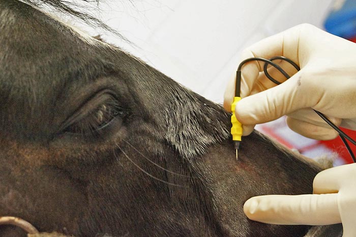
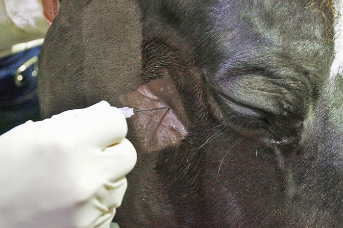
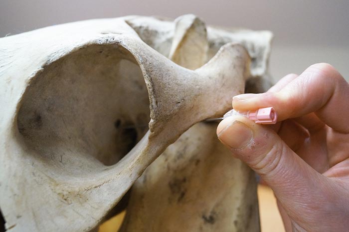
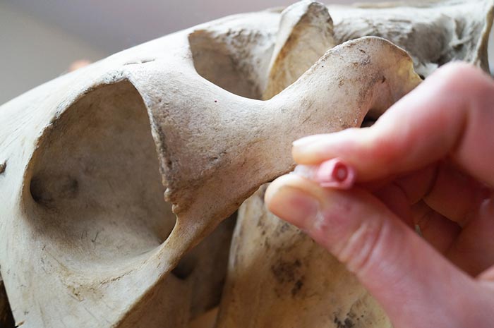
Percutaneous electrical nerve stimulation (PENS) therapy has been proposed as a repeatable, minimally invasive neuromodulatory therapy for the management of TMH24. The technique is effective in reducing trigeminal and other neuropathic pain in humans25. A disposable probe is placed SC to overlay the infraorbital nerve as it exits the infraorbital foramen (Figures 5 and 6).
An alternating current is then applied and the voltage increased to the maximum level the horse will tolerate. A single report of the technique has been published in which five of seven horses improved; the period of remission was variable from 0 to 28 weeks24. More recent follow-up on around 80 horses is less promising, with approximately 25% of horses responding for greater than two months after a course of treatment, approximately 25% responding for less than two months and 50% not responding (Veronica Roberts, personal communication).
The procedure is safe. Haematoma formation is a common complication of probe placement, but is self-limiting and some horses exhibit more severe clinical signs for a few days following the procedure. Serious or long-term adverse effects have not been reported. Predictably, the technique does cause considerable discomfort in some horses as the nerve is stimulated and when the nerve is touched during probe placement.
Infraorbital neurectomy5 and posterior ethmoidal nerve sclerosis are not recommended as a high rate of severe and irreversible adverse effects exists3. The implantation of platinum coils into the infraorbital canal was proposed as an alternative to neurectomy and has been associated with long-term improvement in 50% of cases13,26; however, it has fallen out of favour as most horses undergoing the procedure develop severe discomfort and self-traumatise following surgery.
Recurrence of clinical signs after the first procedure was common. In most horses, the adverse effects were short-term (days), but in some, they were permanent and euthanasia was indicated.
Most horses that exhibit signs of headshaking have reduced activation thresholds of their trigeminal nerves. The cause is unknown. Diagnosis of TMH is by exclusion of other potential causes of headshaking.
The quality of evidence for virtually all treatment interventions is extremely poor and controlled blinded investigations are badly needed. As things stand, none of the treatments proposed for the condition perform better than a placebo.