19 Jun 2023
Kayleigh Cook BVSc, CertAVP(ESST), MRCVS outlines the various forms of this common skin neoplasia and some of the treatments under development.
Kayleigh Cook
Job Title
Image: © callipso88 / Adobe Stock
The most common site of neoplasia in the horse is the skin, and equine sarcoids are widely considered to be the most common form of cutaneous neoplasia (Hollis, 2023; Scott and Miller Jr, 2011).
Sarcoids are locally invasive fibroblastic tumours and their growth has been associated with infection with bovine papillomavirus (BPV) types 1, 2 and 13 (Hollis, 2023; Lunardi et al, 2013).
Vectors such as flies have been implicated, but not proven, as one potential transmission pathway of these viruses (Finlay et al, 2009). Genetic factors have also been implicated in the pathogenesis of the condition (Scott and Miller Jr, 2011).
Although sarcoids do not metastasise, they can become a major welfare concern in affected individuals, as well as leading to a loss in value of the animal (Hollis, 2023).
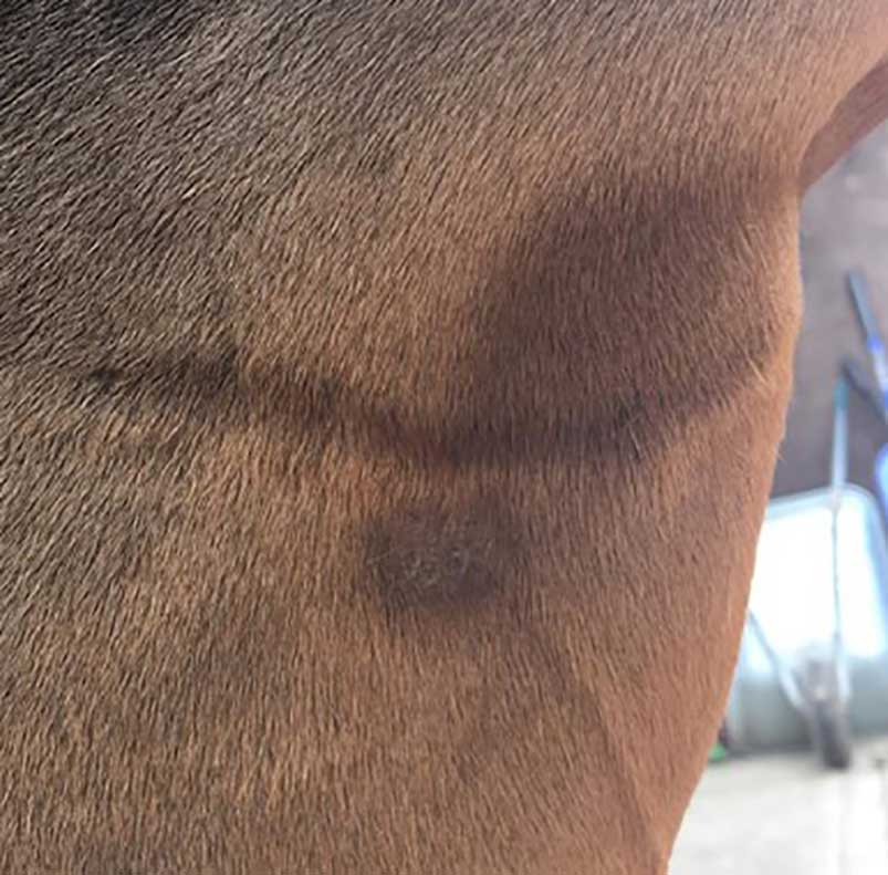
Sarcoids most commonly develop in horses between the ages of one to seven years old. No specific predilection sites exist as they can develop anywhere on the body, with many horses having multiple lesions. They are most commonly found on the head, neck, limbs and ventrum, and are often in close association with a blood vessel (Figure 1; Scott and Miller Jr, 2011). Sarcoids have been noted to develop more commonly at sites of previous trauma or injury (Koch et al, 2018).
Six broad categories of sarcoids are recognised based on how they appear clinically: occult, verrucose, nodular, fibroblastic, mixed, and malignant (Knottenbelt, 2005, Knottenbelt and Kelly, 2000). Many lesions present with features of more than one type, so this is not a strict classification (Hollis, 2023).
Occult sarcoids (Figure 1) are the most superficial and generally considered the mildest type of sarcoid. They are commonly found in sites of minimal hair, such as peri-orbitally and on the inner thigh.
Early lesions can be subtle with hair loss and mild hyperkeratosis, often progressing to contain small nodules in the later stages. Some horses seem to be affected by large numbers of these lesions (Knottenbelt, 2005).
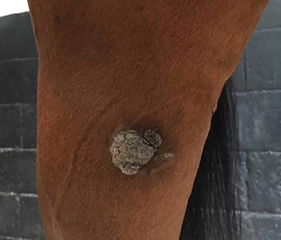
Verrucose sarcoids (Figure 2) have a rough, thickened and scaly appearance, often appearing as wart-like growths. This type of sarcoid is found most commonly on the face, axilla and groin, and can cause significant functional effects on the upper eyelid when they occur in this location (Knottenbelt, 2005; Knottenbelt and Kelly, 2000).
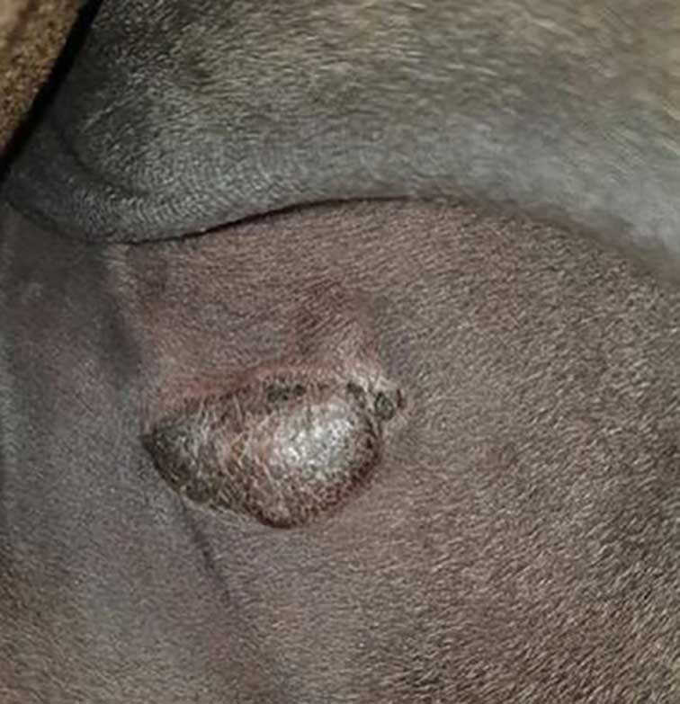
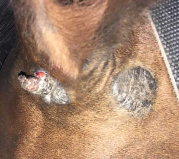
Nodular sarcoids (Figure 3) are round, well-defined subcutaneous nodules and can vary in size from a few millimetres to several centimetres in diameter. They can occur singly as pea-like lumps or in a cluster of interconnecting masses.
Two distinct forms have been noted to occur: type A and type B nodules. Type A have no dermal attachment. Type A1 nodules can move independently of overlying skin and underlying subcutaneous tissue; type A2 nodules have some deeper attachment to underlying layers.
Type B nodules have dermal involvement. Type B1 nodules only have an attachment to overlying skin; type B2 nodules have both skin and subcutaneous tissue attachment (Hollis, 2023; Knottenbelt, 2005).
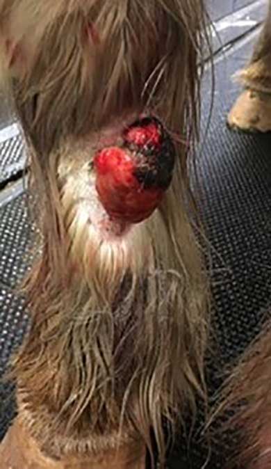
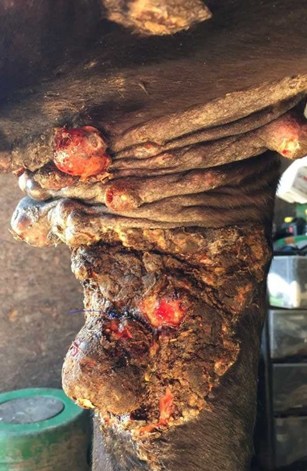
Fibroblastic sarcoids (Figure 4) have an ulcerated and aggressive appearance, and in some instances, can resemble exuberant granulation tissue or “proud flesh”. They tend to appear most commonly on the ventrum, limbs and at sites of previous trauma.
Two forms of this sarcoid type exist: type 1 (pedunculate), which can be separated into type 1A and 1B, and type 2 (sessile). Type 1A growths have a narrow pedicle with no significant root. Type 1B sarcoids have a pedicle, but with a significant deep base at the root, which can make treatment more difficult.
Type 2 lesions have a broad, locally invasive base, and may represent a progression of an initial lesion following trauma or partial treatment.
As all forms of this sarcoid type can resemble exuberant granulation tissue, it is worth considering this as a potential cause of delayed wound healing in a chronic wound.
Histological analysis of excised granulation tissue incurs minimal cost and may reveal the presence of sarcoid – therefore, altering the treatment plan (Knottenbelt, 2005).
Mixed sarcoids (Figure 5) may represent the later stages of a sarcoid transforming from an occult to a verrucose form, or from a nodular to a fibroblastic form. In clinical cases, most sarcoids possess the characteristics of multiple forms of sarcoids, and so, would likely fall into this category (Knottenbelt, 2005).
Malignant or malevolent sarcoids (Figure 6) occur most commonly on the face or upper limbs. Any trauma to a sarcoid, ranging from contact abrasion from tack to partial excision, makes transformation to a more aggressive form more likely.
Some sarcoids of this type can infiltrate lymphatic vessels, forming cords of tumours away from the central mass (Knottenbelt, 2005).
Definitive diagnoses of sarcoids rely on histological examination of a biopsy taken from a suggestive site. However, this is rarely performed in practice due to concerns over causing trauma to the sarcoid and increasing the chance of aggressive transformation (Knottenbelt, 2005; Koch et al, 2018).
Therefore, diagnosis is commonly based on a high index of clinical suspicion given the signalment and presentation of lesions.
Studies have shown that a diagnosis based on clinical presentation alone is accurate in 82% of cases, with a sensitivity of 83.3% and a specificity of 79.6% (Koch, et al, 2018). Therefore, it is reasonable to consider treatment of sarcoids without definitive histological confirmation when clinical appearance is heavily supportive of the diagnosis.
Biopsy should be reserved for cases with a higher degree of uncertainty, and owners should be counselled on the need for further treatment if histopathology is consistent with a sarcoid (Knottenbelt, 2005; Koch et al, 2018; Taylor and Haldorson, 2013).
Several treatment options exist for equine sarcoids. All options have a variable success rate, with none providing a uniformly effective remedy for all sarcoids in all locations (Taylor and Haldorson, 2013).
Benign neglect is rarely advised as a treatment option. Sarcoids are a neoplastic transformation; therefore, it is advised that all cases are treated as early as possible in the course of the disease to achieve the best outcome (Hollis, 2023; Knottenbelt, 2005). This is particularly vital for sarcoids in areas such as peri-orbitally, where a high risk exists of reduced functional outcome if the mass becomes invasive (Knottenbelt and Kelly, 2000).
Some studies report spontaneous regression of sarcoids, although it is uncertain how commonly this phenomenon occurs, and so, it should not be relied on (Martens et al, 2001).
Ligation of sarcoids is a widely used treatment method that can work well on those masses without a significant base. It involves removing the blood supply to a growth by placement of an elastic ligature at the base.
This method is not without risks and should never be used on sarcoids with suspicion of deeper involvement. Combining this with adjunctive methods to ensure no residual cells remain at the base of the sarcoid is advised (Knottenbelt, 2019).
Traditional surgical excision can be used to treat sarcoids in accessible areas.
Wide margins of excision of 2cm to 3cm should be used to ensure removal of the entirety of the sarcoid margin (McCauley et al, 2002). Any masses that regrow following this form of excision are likely to be invasive, and so, should be treated promptly if they occur.
Success rates for this treatment have been reported at 30% to 50%, with regrowth of most lesions being seen by six months – particularly in areas such as around the eye (Knottenbelt and Kelly, 2000; McCauley et al, 2002).
Adjunctive treatments provided alongside conventional sharp excision may improve success rates for this treatment method (Hollis, 2023).
Surgical removal of sarcoids with the use of diode or CO2 lasers offers some advantages over traditional surgical excision.
They vaporise soft tissue as they cut, which reduces intraoperative haemorrhage, lowers the risk of seeding of sarcoid cells, and results in a reduced degree of postoperative swelling compared to sharp excision (Palmer, 1996).
Success rates of 62% to 83% have been reported using this technique (Compston et al, 2016; McCauley et al, 2002). It is thought that excision with diode lasers increases the margins of excision, as it tends to lead to a wider zone of thermal necrosis compared to a CO2 laser and, therefore, provides a reduced chance of sarcoid recurrence (Hollis, 2023).
Electrosurgery is a technique performed under general anaesthesia, with reported success rates of 86.8% in one study. This study used 12mm margins of visibly normal skin and a non-touch technique alongside frequent glove and instrument changes (Haspeslagh et al, 2016).
Cryotherapy can be used as a sole therapy or following surgical debulking as an adjunctive therapy. Liquid nitrogen at -196°C is applied via a spray or probe to the site, leading to intracellular ice formation and, therefore, rupture of cell membranes.
Sloughing of tissue occurs in two to four weeks and tissue depigmentation at the site may remain for months following treatment (Hollis, 2023). Repeated treatments may be required, and severe scarring can occur with this treatment method; therefore, it is not advised for the treatment of periocular sarcoids (Knottenbelt and Kelly, 2000).
Overall success rates for this modality following surgical debulking are reported to be 70% to 80% (Martens et al, 2001).
Radiotherapy is available in three main forms: teletherapy, strontium plesiotherapy and brachytherapy.
The technique utilises ionising radiation to destroy neoplastic cell DNA and proteins (Hollis, 2023; Taylor and Haldorson, 2013). It is considered a gold standard treatment for periocular sarcoids; however, its use is limited by costs and availability of treatment centres (Knottenbelt and Kelly, 2000).
Teletherapy involves the use of high-energy radiation from a linear accelerator being applied to a tumour under general anaesthesia over several treatment sessions. Brachytherapy involves placement of a small, sealed radioactive source within a tumour for up to three weeks (Taylor and Haldorson, 2013).
This treatment is used less often these days due to practicalities, and health and safety issues. A high-dose brachytherapy technique has been used recently, which is performed under standing sedation and has reported good success rates.
Strontium plesiotherapy is a technique suitable for use in superficial lesions. It can be performed under standing sedation without the need for isolation of the horse and appears to be effective in a small number of case reports (Hollis, 2023).
Radiotherapeutic techniques yield success rates of 74% to 94% in various studies (Byam-Cook et al, 2006).
Intralesional cisplatin can be injected into masses or implanted in the form of cisplatin beads, with variable success rates of 33% to 96% reported (Haspeslagh et al, 2016; Hollis, 2023; Knottenbelt and Kelly, 2000).
Intralesional mitomycin C, 5-fluorouracil, carboplatin and bleomycin have all been described with variable success rates. All forms of this treatment impose important health and safety consideration for all handlers of the horse, so their use should be considered carefully (Hollis, 2023).
Electrochemotherapy is a novel technique for sarcoid treatment that uses electrical field pulses to increase cisplatin delivery to a tumour by increasing cell membrane permeability.
It requires repeated treatments under general anaesthetic, depending on the size of the mass being treated (Taylor and Haldorson, 2013). It can be used alone in combination with surgical excision, with reported success rates of up to 98% (Tamzali et al, 2012).
Topical therapeutic options are often considered a low-cost and easy treatment option for sarcoid treatment. However, a few downsides to their use exist.
Transfer of applied material to unaffected surrounding skin is a frequent occurrence, despite reasonable precautions being taken. In some cases, this can lead to significant irritation and pain.
Accidental exposure to handlers is also increased if material is rubbed off on to the environment. Topical application leads to a variable uptake of chemotherapeutic agent into the lesion.
Repeated applications and treatment courses are often required and can lead to significant scarring.
5-fluorouracil has been used topically, with variable success rates of 26.7% to 77% (Hollis, 2023; Knottenbelt and Kelly, 2000). AW3, AW4 and AW5 cream has been used for some years, with anecdotal reports of good success.
Several treatments must be applied, and marked local oedema and pain is often seen because of treatment.
Large open wounds can be left once necrotic tissue is sloughed off, which can require a significant period to heal.
Other topical therapies, such as imiquimod, bloodroot ointment, acyclovir and tazarotene have all been used in the treatment of sarcoids with variable anecdotal success rates, but with minimal controlled studies to assess efficacy (Hollis, 2023).
Immunotherapy has previously been a commonly used form of treatment for sarcoids. Intralesional injection with Bacillus Calmette-Guérin (BCG), an attenuated strain of Mycobacterium bovis, stimulates a local cell mediated immune response, leading to elimination of tumour cells.
Success rates have been reported at 58% to 69%. Significant risk of anaphylaxis exists with this treatment method, which should be considered before use. Premedication with anti-inflammatories may reduce this risk.
Recently, BCG has become more difficult to obtain, which has limited its use (Hollis, 2023).
Recent research has worked on the development of vaccinations made up of chimeric virus-like particles that cause tumour regression. Reports have shown successful use in around 50% of cases (Taylor and Haldorson, 2013).
However, BPV has not been ubiquitously proven as a cause of sarcoids, so this adds some difficulty to the formation of vaccine. As flies are implicated in the transmission cycle of sarcoids, vaccination would need to be undertaken at a very young age prior to exposure of horses to flies, which may not be feasible (Hollis, 2023).
Sarcoids are a frequent and constant challenge to both horse owners and equine veterinarians.
Much still exists that we do not understand about the pathogenesis and transmission cycle of sarcoids, which limits us in our ability to effectively prevent and treat this disease.
Despite the wide array of treatments available for sarcoids, we lack one singularly effective treatment method.
It is clear this is an area that requires further research to improve the health and welfare of horses worldwide.
Use of some of the drugs mentioned in this article is under the veterinary medicine cascade.