30 Oct 2018
Patrick Pollock discusses how advances in imaging and overground endoscopy have improved the understanding of this issue, and how more efficacious therapies have enhanced horse welfare.
Patrick Pollock
Job Title
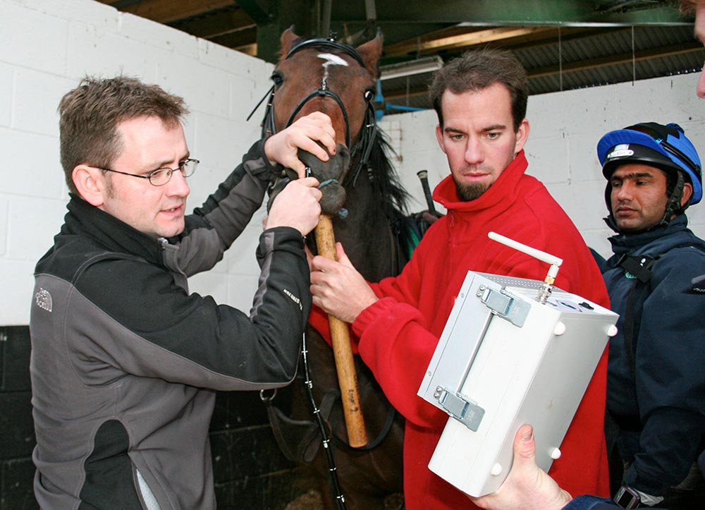
A horse undergoing overground endoscopy with an early version of the equipment under normal exercising conditions at home.
The unique arrangement of the upper respiratory tract (URT) – unsupported by bone or cartilage for much of its length – and the equine requirement for nasal breathing leave horses vulnerable to any disease or dysfunction, resulting in narrowing of the airway.
To date, a great variety of obstructive diseases of the upper airway have been described. However, diagnosis may be complicated by the fact a significant proportion of these diseases only occur during high-speed exercise.
It is fair to say the history of therapy for conditions affecting the URT has not always been based on evidence. The pressure on veterinary surgeons, from owners and trainers of horses, to intervene when presented with a poorly performing horse is often immense.
As a profession, we have a history of selecting a course of therapy for horses with apparent airway problems based on the presence of airway noise (Burn and Franklin, 2006), resting endoscopic findings, the age of the horse or because it is what has always been done on a particular premises (Cheetham et al, 2008; Reardon et al, 2008). The scientific basis for therapy has been further damaged by the, often outspoken, opinions of well-known trainers and veterinary surgeons.
The historic approach to the investigation and treatment of the equine airway has, undoubtedly, not been the profession’s finest hour. Thankfully, this has started to change because, with the use of overground endoscopy, the surgeon has nowhere to hide.
It is now possible for every horse to be examined undertaking the type and intensity of exercise in which it reportedly has a problem. Subsequently, no horse should be treated without a diagnosis. In some cases, that may even involve examining the horse on a racetrack, with other horses, and performing under race conditions. Following the acquisition of the endoscopic video, a diagnosis can be made and an appropriate therapy selected.
In the author’s clinic, all horses presented for overground endoscopy that have a subsequent surgical treatment are offered a free repeat overground examination following their return to exercise. Consequently, the vast majority of procedures are audited, allowing a great deal of evidence to be collected. In days gone by, the only measure of success was performance, and no doubt exists that performance is a poor index of surgical success.
Any horse presented with a history of poor performance should be subjected to a full clinical examination. Although respiratory causes of poor performance are common, dysfunction of the musculoskeletal system still accounts for the greatest number of veterinary call-outs to underperforming horses.
Consideration should be given to the history:
Vets must remain open to the idea poor performance can be related to lack of ability, rather than a clear veterinary problem – and horses affected in this way can present an enormous challenge.
Endoscopy of the URT at rest is performed early in the examination, and provides information on the structure and function of the airway.
This examination should be performed with a video endoscope passed – where possible – through the same nostril, in the correct orientation and for sufficient time to allow detection of any abnormalities present at rest. Sedation should be avoided due to the potential to exacerbate or improve the appearance of some abnormalities.
The diagnostic potential of resting endoscopy may be improved by performing the examination before and after exercise, and by nasal occlusion. Many abnormalities of the airway can be identified using this technique, although it is now accepted resting endoscopic examination poorly reflects the appearance of the airway during high-speed exercise (Kannegieter and Dore, 1995; Tan et al, 2005; Lane et al, 2006b).
The fact many abnormalities of the upper portion of the airway of horses occur together further highlights the importance of overground respiratory tract evaluation (Lane et al, 2006a).
The use of a single resting endoscopic examination early in an animal’s life, as a measure of future performance, has been called into question. No doubt exists the resting appearance of almost all structures of the pharynx, except the arytenoids, is of little value in predicting future athletic success. Horses with evidence of laryngeal asymmetry grades 3 out of 4, or greater, using the Havemeyer scale, have been shown to have reduced earnings and shorter careers when compared to horses graded 1 or 2 out of 4.
Overground endoscopic examination of the URT is one of the most exciting innovations in veterinary medicine. The equipment, of which a number of versions are commercially available, allows the acquisition of images of the URT under normal exercise conditions and does not require the use of a high-speed treadmill (Franklin et al, 2008; Pollock et al, 2009).
With the widespread use of the technique, availability of equipment and the expertise to use it, a giant leap forward has occurred in the understanding of the structure and function of the equine upper airway, and, consequently, a paradigm shift in the approach to – and therapy for – diseases of the equine airway.
Before the introduction of overground endoscopy, high-speed treadmill endoscopy was considered the gold standard for diagnosis of diseases of the URT. However, the lack of rider and tack; difference in surface, speed, incline and environment; potential difference in head position; and lack of other horses has led workers to conclude treadmill endoscopy poorly emulates normal exercising conditions (Evans, 2004; Franklin et al, 2008; Pollock et al, 2009).
Most vets involved in the development of overground endoscopy expected a system allowing examination of horses in their environment, and in larger numbers than those presented for treadmill investigation, would result in greater numbers of airway surgeries for clinics that regularly treated horses. This has not been the case. Instead, the technique has allowed clinicians to better understand the airway, monitor the development and progress of disease, and – perhaps most importantly – assess its significance on the exercising horse. From a welfare perspective, little doubt exists the single biggest contribution overground endoscopy has made is a reduction in the number of horses subject to surgical procedures of dubious efficacy.
To date, the author’s clinic has performed about 5,000 overground examinations, with many more undertaken around the world. The technique is straightforward, reliable and safe. Breeds examined have included Thoroughbreds, Thoroughbred crosses, standardbreds, sport horses, Clydesdales and a number of native-breed ponies.
In the population of horses scoped to date, a number of pathological conditions have been identified, including:
A number of new conditions have also been described (Pollock et al, 2013).
Diagnosis of these conditions, many of which were only visible during strenuous exercise, is encouraging. Many questions exist about their significance, with reference to effect on performance, links to abnormal respiratory noise and repeatability of diagnosis. It has rapidly become clear a considerable amount of work is required in investigating upper airway pathology in ridden horses under normal exercise conditions.
The system has resulted in the presentation of horses not traditionally or regularly presented for treadmill examination, and to the representation and serial examination of horses after tack changes, or conservative or surgical treatment. It requires few operators and does not require hospitalisation, so is considerably cheaper than treadmill endoscopy.
Examination of the equine airway is further complicated by the fact the appearance of the pharynx and, particularly, the larynx of the same horse may differ from hour to hour, or day to day.
This variation in laryngeal symmetry has been demonstrated during resting and overground endoscopy. Consequently, if the endoscopic appearance of the airway does not fit the clinical signs or history, repeat examination is warranted, and serial examinations may be required before a diagnosis can be made and an appropriate treatment selected.
An increasing number of scientific studies have been performed using overground endoscopy, some of which have given rise to changes in the approach to treatment.
In a group of 100 racehorses with a diagnosis of obstructive disease of the upper airway, presented due to the presence of abnormal respiratory noise, a comparison of recorded noise and overground endoscopy findings suggested use of respiratory noise alone as a diagnostic criteria would result in an incorrect diagnosis in approximately 50% of horses presented (Witte et al, 2011). In a significant proportion of horses in this study, abnormal noises were recorded in horses with no evidence of airway disease.
Longitudinal studies have suggested overground endoscopy in young horses can identify individuals that may develop significant and career-limiting airway problems (Kelly et al, 2013).
Conservative therapy for equine airway problems have never been all that popular; the pressure on vets to “do something” has often led to the use of surgical treatments that are at best ineffective and at worst damaging. This is particularly true for treatment of intermittent dorsal displacement of the soft palate.
Due to the ease that overground endoscopy can be performed, it has been possible to serially examine large groups of horses with this condition. The findings suggest, in young horses, this condition is likely to be self-limiting in the majority of cases (more than 70%).
Despite the rise in conservative therapy for a number of common URT problems, instances still exist when surgical intervention is justified.
No area is developing as quickly as treatment of recurrent laryngeal neuropathy. Traditional treatment of this condition still includes removal of the laryngeal ventricles and one or both vocal cords, and a prosthetic laryngoplasty; however, the redevelopment of laryngeal reinnervation techniques – that allow the paralysed cricoarytenoid dorsalis muscle to regenerate – mark the next exciting step in the science of the structure function and treatment of the equine upper airway.