17 Oct 2016
Andy Durham outlines some diagnostic and treatment approaches to ulceration of the cornea presenting in horses.
Andy Durham
Job Title
Figure 2c. An ulcer demonstrating evidence of acute necrosis (“melting”).
Corneal ulceration is a common problem in equine practice. Horses are inquisitive animals and have prominent eyes – a combination making them predisposed to traumatic injury of the cornea.
The majority of cases are simple and respond well to empirical treatment, but, unfortunately, many cases are also encountered that stubbornly refuse to heal. This article intends to outline a diagnostic and treatment approach to such cases.


Examination of painful equine eyes is an almost impossible procedure unless the horse is sedated well and an auriculopalpebral nerve block is performed. Important findings, such as foreign bodies and conjunctival nodules, are easily missed, as are ulcers themselves, unless proper control of the horse and its eyelid is achieved.
The auriculopalpebral branch of the facial nerve (CrN VII) controls the muscle of the upper eyelid and is blocked where it can usually be palpated subcutaneously running over the zygomatic arch just caudal to the ventral depression of the arch, caudal to the orbit (Figure 1).
Between 2ml and 5ml of local anaesthetic is used via a 25-gauge needle. The eye should then be carefully and closely examined with direct illumination, with you looking closely for irregularities in the corneal surface, grey corneal oedema, vascularisation and any evidence of secondary uveitis (especially a constricted pupil). If any ulcers are found then the overlying conjunctiva should be carefully checked for irregularities or foreign bodies.
Fluorescein dye can be used to better demonstrate ulcerated epithelium, but should be diluted, rather than applied as a fluorescein strip directly to the eye, which tends to introduce far too much dye. The author prefers to place a fluorescein strip into a plain vacutainer containing 5ml sterile water and then using this diluted fluorescein in the eye itself via a 2ml syringe and needle hub after breaking off the needle. Corneal stromal staining is massively enhanced by shining a blue light on to the eye in a darkened stable.
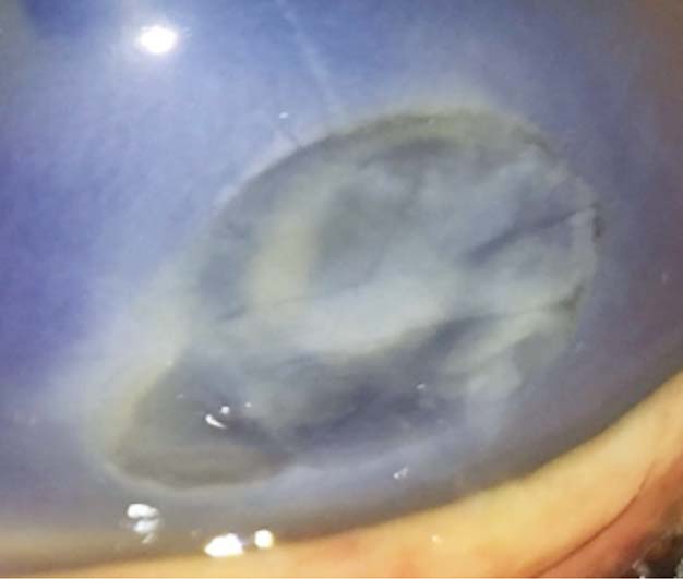
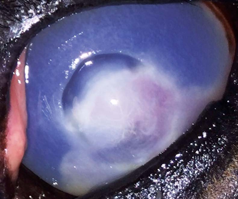
A general simple assessment of the nature and depth of the ulcer is important prognostically (Figure 2). A simple superficial corneal deficit is encouraging, although does not mean it will inevitably heal quickly. A deeper ulcer continuing into the corneal stroma will inevitably take longer to heal.
Evidence of necrosis with a mucoid, mushy cornea is certainly alarming and indicates a more aggressive, urgent and possibly surgical approach is required.


Sampling from the ulcer is important – especially if concerning features are present, such as marked uveitis, stromal involvement or a failure to respond well to initial medication. A smear collected from the ulcerated area can offer crucial clinical information within minutes. It is better to collect this using the blunt edge of a scalpel blade (or a cytology brush) rather than a swab as the possible detection of fungal hyphae in the smear cannot then be confused with filaments from the swab.
Early diagnosis and specific treatment of fungal keratitis is very important and not uncommon – especially in the warmer months. Additionally, especially problematic pathogens, such as streptococci, can be immediately identified in the stained smear as chains of cocci. A swab can then be collected following the smear for further culture (bacterial and fungal), although this will inevitably take a few more days.
Bacterial infection of the ulcer warrants topical antimicrobial therapy, although it is important not to be too led by the reported antimicrobial sensitivity patterns reported by the laboratory. Predicted sensitivity/resistance and also minimum inhibitory concentration (MIC) values are designed with reference to predicted systemic drug levels and have little relevance to the far higher concentrations achieved in tear film with topical ophthalmic preparations.

For example, Enterobacteriaceae or Pseudomonas with an MIC of 16µg/ml would be reported as resistant based on expected maximum plasma drug concentrations following IV gentamicin of perhaps 40µg/ml (that is, less than 8 × MIC).
However, a good clinical response to topical gentamicin drops is very likely as it is known just three drops of 0.5% gentamicin can achieve greater than 350µg/ml in tear film, high enough to kill even apparent highly resistant bacteria. Voriconazole is generally the preferred topical antifungal agent, although it is expensive and often not readily available. Where it is not available, topical enilconazole (diluted 1 in 50) can be used or, alternatively, clotrimazole ointment.
Where ulcers are showing slow or absent healing then consideration should be given to blocking the effect of matrix metalloproteinases, which impede healing. Ulcerated corneas contain at least three times normal matrix metalloproteinases (MMP) concentrations in the tear film and, interestingly, the contralateral normal eye also tends to have higher MMP levels than normal, suggesting a partially systemic effect on corneal MMP expression.
Many choices of antiproteinase exist, including autologous plasma, serum, 20% acetyl cysteine and calcium chelators, such as 0.2% ethylenediaminetetraacetic acid (EDTA) solution and tetracycline ointment. All have been shown to be effective although some are more active against specific MMP isotypes, meaning at least two different agents may be indicated. Studies have indicated serum and doxycycline have better activity versus MMP-2 whereas EDTA and acetylcysteine have better activity against MMP-9. It is the author’s general practice to alternate antiproteinase treatments with autologous serum (or plasma) and 0.2% EDTA solution with the intention of broad-spectrum antiproteinase effects.

The timing and duration of antiproteinase treatment is perhaps the most crucial element of treatment of non-healing corneal ulcers and, in this author’s view, probably the primary reason for treatment failure. As aforementioned, MMP concentrations are increased in tear film of ulcerated eyes and intuitively will constantly resist healing and/or promote deterioration.
It should be considered any agent instilled into the eye is unlikely to remain present for longer than 10 or 15 minutes before being washed away by the tear fluid. Thus, even given an example of application of antiproteinase every four hours (which would require hospitalisation or a very committed client) then the MMPs are very unlikely to be inhibited for longer than 6% of the time (that is, 6 × 15 minutes per 24 hours), leaving them uninhibited and attacking the cornea still for the other 94% of the time.
Even hourly instillation would only provide 25% MMP inhibition over time (24 × 15 minutes per 24 hours). Thus, serious thought should be given to continuous infusion of antiproteinases via a subpalpebral catheter, making use of a balloon reservoir or infusion pump device. Unfortunately, this generally means hospitalisation is required, although the increased chances of achieving reasonably prompt healing will often make this a cost-effective option.
Indeed, subpalpebral lavage catheter placement is pretty much mandatory for any horse (however good natured) receiving intraocular medications more than three or four times daily. Catheters are easily placed under sedation, with the lower lid towards the medial canthus being the preferred site to avoid complications. Lower eyelid subpalpebral lavage catheters are extremely well-tolerated, with complications being exceedingly rare (Figure 4).
Additional relatively simple tactics that can be used alongside aforementioned medications include grid keratotomy, diamond burr debridement and tarsorrhaphy. Grid keratotomy can be performed under sedation and topical analgesia of the cornea. Although the equine cornea is less than 1mm thick, it is a pretty tough structure and light application of the edge of a 19-gauge needle, a scalpel blade or a specific depth-limited keratotomy knife, is a very low risk procedure in sedated horses.
Published studies indicate good resolution rates of non-healing ulcers post-keratotomy (for example, 70% healed within two weeks), although the author’s experiences have not been quite so dramatic. Similarly, diamond burr debridement (Figure 5) can be applied to corneas after sedation and topical analgesia, and this carries an even more impressive published healing rate of greater than 90% at two weeks.

Again, however, although a probably useful technique, the author’s experiences have been frequently disappointing following this procedure. As long as the non-healing ulcer appears stable – at least with no obvious threat of deeper extension, abscessation or uveitis – then tarsorrhaphy should also be considered. The author has frequently found chronic non-healing ulcers heal well during a week of enforced eyelid closure (perhaps due to absence of frictional forces on the delicate early healing ulcerated epithelium during blinking).
This can be achieved with one or two sutures carefully and gently placed from the upper to lower eyelids. Ultrasound can be used to offer reassurance of globe integrity, corneal thickness, pupil size, aqueous clarity and so on during the period of tarsorrhaphy – although, as aforementioned, this technique is only really indicated in ulcerated corneas that appear stable and uncomplicated, albeit non-healing.
Finally, surgical options should be considered either when conservative medical approaches have failed after several weeks of treatment, when further threat to the eye is seen, such as progressive deepening or necrosis of the ulcer, or as a means of attempting to expedite a slowly healing corneal ulcer. Conjunctival pedicle flaps may be placed under general anaesthesia and offer both structural stability and vascularisation of the ulcer bed.
Provided dehiscence does not occur then good strength and healing is expected within about two weeks of surgery. Permanent scarring of the ulcerated area is to be expected, which could cause significant visual deficit if large. Application of amnion grafts tends to encourage corneal healing with much less, if any, residual scarring, but it is offered by relatively few equine hospitals in the UK.
Non-healing corneal ulcers are among the more frustrating conditions seen in equine practice and clinicians should be forgiven for feeling like innocent bystanders rather than successful interventionists while watching the horse’s eye making its own mind up whether to heal.
Nevertheless, the aforementioned tactics will improve healing rates in those ulcers that stubbornly refuse to heal – leading eventually to a favourable outcome in the majority of cases.