4 Sept 2017
Sue Dyson reviews literature related to aspects of this issue, in light of clinical experiences and limited evidence-based data.
Sue Dyson
Job Title
It is reasonable to assume biomechanical loading of limbs is influenced by both conformation and the position of the limbs during the stance phase of the stride. Thus, static and dynamic conformation – such as the degree of extension of the metatarsophalangeal joints – could potentially influence the development of lameness.
It also seems logical a well-conformed horse should be at less risk of injury, compared to a poorly conformed horse. However, remarkably little evidence-based data exists concerning conformation and lameness in sports horses.
The purpose of this article, therefore, is to critically review the literature related to some aspects of hindlimb conformation.
A study of 356 Swedish Warmblood horses, judged to be sound based on subjective assessment trotting in hand, found a remarkable variation in hock angles (Holmström et al, 1990).
Four-year-old potential competition horses had a mean hock angle of 155.4° plus or minus standard deviation (SD) 2.6 (range 145° to 161°), whereas the mean values for elite dressage and showjumping horses were 159.1° plus or minus SD 3 (range 150° to 165°) and 159° plus or minus SD 2.7 (range 156° to 165°), respectively. Riding school horses had a mean value of 156.8° plus or minus SD 3.5 (range 148° to 169°).
However, given the high frequency of occurrence of hindlimb lameness not apparent in hand, but obvious on the lunge or ridden (Dyson and Greve, 2016), the likelihood of this representing a population of horses truly free from lameness seems small.
In a comparison of elite dressage horses (n = 40), elite showjumpers (n = 51), quality-tested four-year-olds (n = 217) and horses with “back problems or lameness” seen at two Swedish clinics (n = 52), the hock angles in lame horses (157°) were “significantly” smaller than the other groups (elite dressage horses 160.4°, showjumpers 159.2° and four-year-olds 159.4°). However, probability values were not reported, nor were the causes of lameness specified (Holmström, 2000).
Elsewhere, comparison of hock angles in sports horses examined in 1996 (n = 75), for which insurance data was available in 1998, revealed no significant difference in hock angles between horses with (n = 25) or without (n = 50) recorded lameness (Holmström, 2000).
In a study of preselected potential competition horses that were three years old (German Warmblood, n = 53; Selle Français, n = 61; pure Spanish, n = 28), breed differences appeared to exist (Barrey et al, 2002). Mean hock angles were 157° plus or minus SD 3.6, 155° plus or minus SD 4.8, and 150° plus or minus SD 3.9, respectively.
The biomechanical effects of hock angle were investigated in a group of mixed breed horses (Andalusian, n = 1; Warmblood, n = 3; Arabian, n = 3; Standardbred, n = 6; Thoroughbred, n = 3), aged 6 to 20 years (Gnagey et al, 2006). The horses were assessed as sound by subjective evaluation in walk and trot only; their work history was not documented. The tarsal angles were classified as small (less than 155.5°, n = 4), intermediate (155.5° to 165.5°, n = 8) or large (greater than 165.5°, n = 4). Force plate data was collected at trot for the right hindlimbs only.
Horses with large standing angles had less flexion and energy absorption at the tarsus during the impact phase than those with intermediate or small angles, and generated less vertical impulse than horses with small standing angles. Horses with intermediate tarsal angles generated larger vertical and propulsive impulses than the other groups.
The authors suggested an optimal range of tarsal angles may exist that maximises the ability to push the horse upwards and forwards. However, the group sizes were unequal – four, eight and four, respectively – and the range of tarsal angle in the intermediate group was large (155.5° to 165.5°). Measurements were only made for the right hindlimb, so the symmetry of movement was unknown. Whether all horses were truly non-lame cannot be determined. Less vertical impulse, documented in the horses with large tarsal angles, may actually reflect subclinical lameness.
An association between large tarsal angles and proximal suspensory desmopathy (PSD) has been described (Dyson, 1994).
In a study of 42 horses with PSD, conformation was assessed subjectively and compared with 50 consecutive horses examined without PSD. Three horses with PSD had large hock angles, while six horses had both large hock angles and hyperextension of the metatarsophalangeal joints. In other words, 21% had abnormal conformation, compared with a large tarsal angle in 2% of the control horses. These observations were not verified by objective measurements and all horses had advanced PSD, with overt lameness apparent in hand.
This association does not imply a causal relationship; in fact, it seems more likely hyperextension of the metatarsophalangeal joints is a consequence of compromised function of the suspensory apparatus.
A relationship between large tarsal angles and increased radiopharmaceutical uptake in the proximoplantar aspect of the third metatarsal bone – in association with PSD – has been observed (Dyson et al, 2007), while a study of 155 horses with PSD managed by neurectomy of the deep branch of the lateral plantar nerve and plantar fasciotomy found 5 of 155 horses had hock angles greater than or equal to 165° (Dyson and Murray, 2012).
It was recommended, based on experience, these horses were not good candidates for surgery due to a high risk of persistent lameness or exacerbation of lameness. Surgery was performed elsewhere, with the horses made available for follow-up – all deteriorated. A total of 78% of the remaining horses with primary PSD alone, and with tarsal angles lower than 165°, returned to full function.
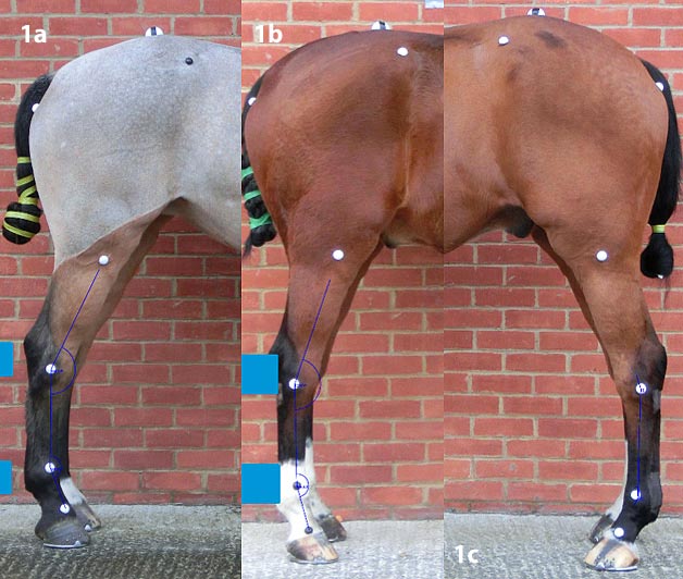
A pilot study comparing tarsal angles of horses of mixed breeds with hindlimb PSD (n = 47; 157.9° plus or minus SD 4.7) and without (n = 18; 154.2° plus or minus SD 3.1), found a difference between the groups of probability lower than 0.001 (Dyson, unpublished data; Figure 1). It is likely, therefore, the aetiology of PSD is multifactorial and conformation is only one component that may represent a risk factor for injury. However, only a long-term, large-scale, longitudinal study could definitively answer whether large tarsal angles predispose to PSD.
In any assessment of hindlimb conformation, it is essential the posture is standardised – horses should be standing squarely on a firm, level surface, with the metatarsal region positioned vertically, and the tuber calcanei and tuber ischii in line.
The relative heights of the withers and tubera sacrale may influence how easily a horse balances itself during locomotion, its ability to engage the hindlimbs and, therefore, the loading on all four limbs.
In Holmström et al (1990) and Barrey et al (2002), the mean height of the withers was greater than the mean height of the tubera sacrale. The maximum and minimum values for the withers were greater than the tubera sacrale, except in elite showjumpers, where the maximum withers and tubera sacrale heights were equal.
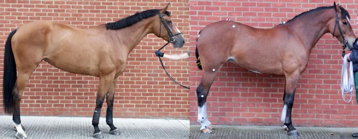
However, in a pilot study of 74 sports horses presented for investigation of lameness or poor performance, based on photographs of horses standing in a standardised position alone, the withers were higher than the tubera sacrale in 66% of cases; the heights were identical in 21%, with the tubera sacrale higher in 13% (Dyson, unpublished data; Figure 2). Comparing this data with Holmström et al (1990) and Barrey et al (2002) suggests the tubera sacrale being higher than the withers may be a risk factor for lameness.
In another study of 296 horses with sacroiliac joint region pain as a contributing factor for poor performance, the tubera sacrale were higher than the withers in 11% of cases (Barstow and Dyson, 2015).
It is important to realise conformation assessed in a standing, static horse does not necessarily accurately predict how the limb will be loaded (“dynamic conformation”) and the influence this may have on injury risk. Plus, no published data exists relating dynamic conformation to biomechanical loading.
It seems intuitive, however, that loading a limb straight, with all the joints in alignment in the sagittal plane, is preferable to the limb being loaded asymmetrically. Sideways oscillation of the hock during the stance phase has been documented in a small number of showjumpers in normal work (Dyson et al, 2016).
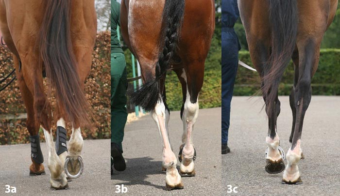
This feature has also been observed in horses with hindlimb lameness (Dyson, unpublished data). It is often associated with the foot being placed further under the trunk than normal, and some rotation of the distal aspect of the limb (Figure 3). Such features are worthy of further investigation with respect to injury risk.
It seems highly likely conformation influences injury risk in sports horses, but remarkably little evidence-based information exists to support this.
Symmetry, proportion and balance are important to assess. Many injuries are likely to be multifactorial in aetiology; therefore, determining the role of conformation – both static and dynamic – is not easy.
The impact of less-than-ideal conformation may be greater in a horse working at the limit of its athletic ability, compared with a horse working well within its capacity. However, a more skilled rider may be able to ride a horse with less-than-ideal conformation better in balance than a less-skilled rider.
Natural extravagance of paces, ground surfaces and work intensity may all play a role. Clearly, many factors need to be considered.