19 Nov 2018
Victoria Colgate and Richard Payne discuss the use of platelet-rich plasma and stem cell therapy in the management of this issue, as well as the need for research into more effective treatments.
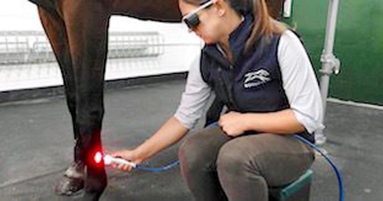
Figure 3. Laser therapy being performed.
Research has highlighted the burden and wastage caused by tendon injuries in equine athletes, yet, so far, failed to undercover a truly efficacious solution.
As the past treatments of counterirritation, tendon splitting and intralesional injection of hyaluronate or polysulphated glycosaminoglycans have largely been replaced by platelet-rich plasma (PRP) and stem cell therapy, so the focus of therapy has shifted from scar formation to regeneration of the naive tendon. However, even these biologics have variable treatment outcomes (PRP), and suffer from technical difficulties in harvesting and preparation of the cells for injection (stem cells).
Through further research, we must better understand the cellular mechanisms of tendon injury to produce a more effective treatment for the future.
Lameness caused by musculoskeletal injury has been identified as a major cause of wastage in the racing industry (Jeffcott et al, 1982; Rossdale et al, 1985; Verheyen and Wood, 2004) – both financially, with days lost training, and in terms of reduced welfare and longevity.
Research has repeatedly highlighted this problem, but failed to reduce injury rates. Development of innovative tendon treatments could have a far-reaching impact, but is complicated by several factors.
The equine superficial digital flexor tendon (SDFT) has a specialised structure allowing storage and release of elastic energy during locomotion (Geburek et al, 2017). Comprised of a high proportion of longitudinally arranged type-one collagen fibrils in an extracellular matrix (ECM; Dowling et al, 2000), the fibrils are further cross-linked to give the tendon its tensile strength (Chesen et al, 2009).
When loaded, fibrils stretch and slide past each other, allowing elongation of up to 20 per cent before failure (Dowling et al, 2000). However, at maximal speeds and loads the SDFT operates close to its physiological limits (O’Meara et al, 2010), predisposing to injury.
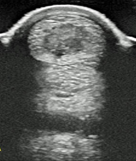
While serious overstrain injuries occur acutely during exercise (Smith and Webbon, 2005), they are thought to be the result of microdamage accumulation and reduced collagen cross-linking (Reed and Leahy, 2013), as repetitive loading during high-intensity exercise superimposes on natural age-related tendon matrix degeneration (Dowling et al, 2000; Geburek et al, 2017; Figure 1).
Intrinsically, the SDFT has “limited regeneration potential” (Romero et al, 2017) and undertakes slow repair dominated by fibroplasia. The resulting hypercellular scar lacks ECM and is largely comprised of disorganised, small-diameter type-three collagen fibrils (Lange-Consiglio et al, 2013). This fibrotically healed tendon lacks elasticity – leading to reduced performance and high risk of re-injury (Godwin et al, 2012).
Treatments have been focused on trying to regenerate naive tendon with strong type-one collagen fibrils, rather than enhancing fibrous repair. The plethora of treatments developed over many decades indicates the lack of a truly efficacious solution.
The conservative approach to tendon repair involves initial box rest, cryotherapy and immobilisation by bandaging, to limit the inflammatory phase of injury where proteolytic enzymes further degrade intact tendon tissue (Dowling et al, 2000). This is followed by a prolonged rehabilitation period of controlled exercise.
As a sole treatment, this produces a slow, inefficient repair (Reed and Leahy, 2013), but the importance of physical loading in ensuring structural realignment of collagen fibrils means controlled exercise remains the cornerstone of treatment, independent of which additional reparative method is used.
Previously, counterirritation by blistering or thermocautery (bar firing or pin firing) was a popular treatment. It had been suggested thermocautery produces thickened, scarified skin to act as a support for the SDFT (Hayward and Adams, 2001), as well as an intense inflammatory reaction within the tendon (Dowling et al, 2000). It has since been found inflammation is deleterious, and with no evidence of a significant effect on tendon healing, counterirritation is, at best, a “brutal means of enforcing rest” (Hayward and Adams, 2001).
Similarly, tendon splitting is thought to release the pressure of haemorrhage and promote revascularisation of the core lesion (Witte et al, 2016). Clinical studies have showed mixed results, and it is argued the surgical trauma created by tendon splitting is at odds with the desired effect (Fackelman, 1973).
Extracorporeal shockwave therapy is thought to assist healing of chronic tendon injuries by stimulating remodelling of scar tissue through increased tenocyte metabolism (Bosch et al, 2009); however, with potential to adversely disrupt normal collagen structure, risk of complete tendon rupture post-treatment has been reported.
Treatments explored so far represent low-cost, minimally invasive options lacking much scientific proof of effect. A greater level of intervention gives a choice between surgical or intralesional treatment, with a number of biologics available.
Desmotomy of the accessory ligament of the affected SDFT is the surgical option. After transection, the accessory ligament heals, but in an elongated fashion – acting to lengthen the overall check ligament-tendon unit (Witte et al, 2011; Hu and Bramlage, 2014). When maximal stress is applied to the affected tendon, this alteration helps negate some of the elasticity lost from the fibrosed tendon repair (Hogan and Bramlage, 1995).
Although success has been reported, this technique has a primary pitfall: lengthening the check ligament-tendon unit helps spare the SDFT, but also transfers weight to the suspensory ligament (Hu and Bramlage, 2014).
With a higher incidence of suspensory ligament desmitis recorded in horses that have undergone check ligament desmotomy (Witte et al, 2016), arguably, the surgical procedure only replaces the existing pathology with a different one.
Intralesional treatments have traditionally involved the injection of hyaluronate or polysulphated glycosaminoglycans (PSGAGs) into affected tendons. PSGAGs are hypothesised to inhibit many catabolic enzymes (Dowling et al, 2000) to improve collagen fibril organisation and increase scar maturation (Moraes et al, 2009), but clinical studies show conflicting results.
β-aminopropionitrile fumarate is a lysyl oxidase inhibitor that delays collagen cross-linking, theoretically allowing time for fibres to align correctly in response to forces experienced during controlled exercise (Dahlgren et al, 2001; Smith et al, 2003).
However, experimental studies have cast doubt on its efficacy and it cannot be recommended for treatment; delaying cross-linking reduces the tensile strength of the healing tendon, increasing risk of catastrophic rupture during early rehabilitation (Dahlgren et al, 2001).
Intralesional injection of corticosteroids – such as methylprednisolone, triamcinolone and dexamethasone phosphate – is contraindicated due to the stimulation of collagen necrosis and hyaluronisation (Dowling et al, 2000).
Most intralesional treatments have been superseded by autologous biologic preparations, such as platelet-rich plasma (PRP) and stem cells. Readily available from patients, with a high safety index, they are not subjected to competition regulations, as occurs with pharmaceuticals (Textor, 2011).
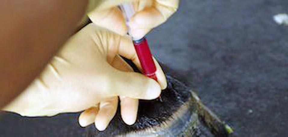
PRP (Figure 2) has been a popular choice for treatment, although supporting evidence for its use is fairly weak (Witte et al, 2016).
PRP is used because platelets contain many growth factors that have an anabolic effect – stimulating tendon healing through enhanced tenocyte proliferation, collagen and matrix synthesis (Geburek et al, 2016).
It is anecdotally noted long-term, PRP treatment has the potential to increase the number of horses reaching their previous performance levels, but in vitro PRP studies have yielded more positive results than clinical trials (Geburek et al, 2016). This is due to the variable nature and composition of PRP, which, as a preparation from an individual horse using a variety of commercial kits, leads to a non-standardised product.
Additionally to platelets, a PRP preparation contains variable numbers of leukocytes that have a proinflammatory effect deleterious to tendon healing (Smith et al, 2014). This has led to the development of autologous conditioned plasma (ACS), which contains fewer white blood cells, therefore creating a lower inflammatory reaction (Georg et al, 2010). Initial studies showed promising results (Georg et al, 2010), but as ACS is thought to have a time-limited effect, it may require repeated injections (Geburek et al, 2015), reducing its feasibility.
With many commercial horse-side filtration systems, PRP is an effective treatment option easily performed in general practice. However, intralesional injection of stem cells is possibly the best regenerative treatment available. Stem cells are precursor cells that have retained the ability to self-renew (Chagastelles and Nardi, 2011) and can be of embryonic or adult origin.
Embryonic stem cells (ESC) are truly pluripotent, capable of differentiating into all adult cell types, derived from any germ layer (Chagastelles and Nardi, 2011). However, while theoretically an ideal therapeutic candidate, ethical issues raised by the destruction of blastocysts in ESC harvesting makes them unfeasible.
Mesenchymal stem cells (MSC) are of adult origin and multipotent, capable of differentiation into cells of a restricted number of tissue lineages (Renzi et al, 2013). Their therapeutic benefit has been clinically proven, but it is unclear whether they have a regenerative effect through direct differentiation into tenocytes or whether healing is stimulated through an indirect paracrine effect (Geburek et al, 2017).
Bone marrow is considered the universal source of MSCs (Shikh Alsook et al, 2015). Cells are harvested from the horse’s sternum and cultured for two to three weeks – to increase cell numbers to 10 million to 20 million (Smith et al, 2014) – before intralesional injection. In the tendon they induce increased type-one collagen expression, allowing regeneration of normal longitudinal fibre pattern (Crovace et al, 2010).
However, timing of MSC therapy is thought to be crucial – with injection needed after early inflammation, but before scar tissue has formed (Romero et al, 2017).
With the three stages of tendon healing (inflammatory, proliferative and remodelling) taking a variable time, it is impossible to predict the perfect time for injection. Additionally, in the three-week delay needed for cell culture, ultrasound changes can occur in the tendon that limit effective regeneration (Lange-Consiglio et al, 2013).
This need for a more immediate – and less expensive – treatment led to the use of bone marrow aspirate as a source of MSCs. The aspirate is harvested and directly injected into the tendon, eliminating the amplification stage. However, it is an unpurified source containing relatively few stem cells (1 × 10⁴ nucleated cells), but with additional fat cells and bone spicules (Smith et al, 2013) that impede healing.
Adipose-derived MSCs are another viable alternative. They are easier to harvest, but less effective (Shikh Alsook et al, 2015), and significant donor site morbidity with seroma formation has been documented (Nixon et al, 2008).
The promise of an effective healing response clinically with use of MSCs has led to further experimental studies to find a more appropriate cell source. Although MSCs are multipotent, they have better regenerative capacity within their tissue of origin (Youngstrom et al, 2016).
Tendon-derived MSCs have been used experimentally to treat SDFT lesions where they have been shown to increase tensile strength and collagen fibre alignment in repaired tissue (Durgam et al, 2016). As these cells have a high proliferation rate, only a small amount of autogenous tendon tissue is harvested to generate clinically useful cell numbers (Durgam et al, 2016). However, morbidity associated with creating a second tendon lesion at the donor site (Alves et al, 2011) is unacceptable, meaning their only potential as a clinical treatment is through allogenic use (Youngstrom et al, 2016).
An allogenic source of stem cells would, in many ways, be ideal, allowing use of unlimited cell numbers from immediately available stock. However, if an allogenic source is to be used, amniotic MSCs probably represent the most exciting treatment prospect for the future.
Lange-Consiglio et al (2013) showed re-injury rate in horses treated with allogenic amniotic membrane-derived stem cells was lower (4 per cent) compared to the average observed with autologous bone marrow-derived MSCs (23 per cent). With amniotic MSCs being more primitive than bone marrow MSCs, they provide potential for better engraftment (Lange-Consiglio et al, 2013) and are available for collection in unlimited numbers from extrafetal tissues discarded at parturition (Lange-Consiglio et al, 2012).
Once storage by cryopreservation has been refined (Lange-Consiglio et al, 2012), potential exists to create a bank of stem cells for future, immediate intralesional use (Lange-Consiglio et al, 2012; Reed and Leahy, 2013).
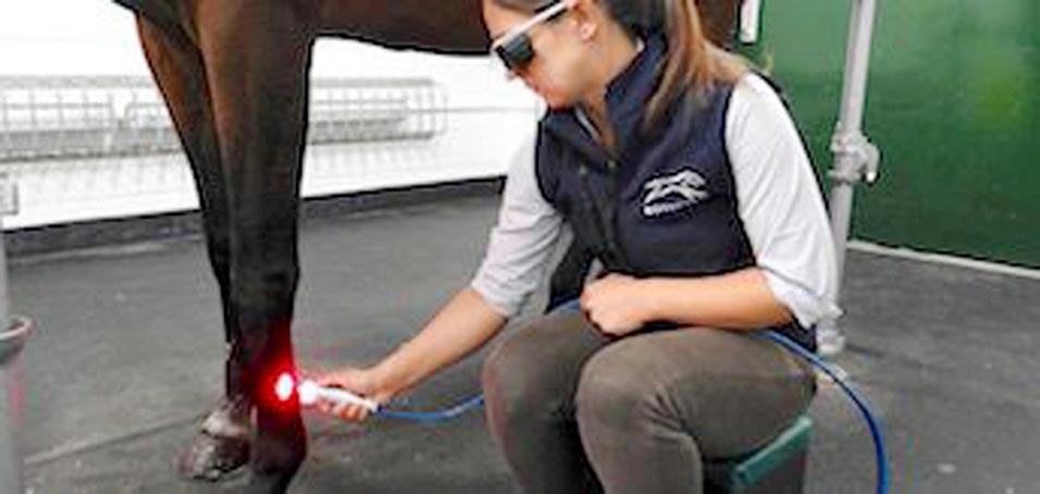
Although technically advantageous, the practicalities of horse-side treatment with stem cells is challenging – and of recent evolutions, laser therapy (Figure 3) may represent the most promising and feasible option.
Anecdotally in the author’s clinic – and as reported by Pluim et al (2018) – it results in a more rapid improvement in clinical lameness and ultrasound score than previously described treatments. While this allows earlier return to work, only time and further studies will elucidate if the tendon repair is as good as its ultrasonographic appearance.
It may seem little progress has been made in tendon treatments over the past few decades, but this reflects the complexity of the problem, which is not restricted to veterinary medicine.
Interestingly, the human Achilles tendon is functionally analogous (Durgam et al, 2016) to the SDFT in horses, and equine SDFT lesions are a well-accepted model of human exercise-induced Achilles tendon injury (Patterson-Kane and Rich, 2014). With the SDFT being the “most completely understood tendon injury in any species” (Patterson-Kane and Rich, 2014), we should not feel disheartened at progress as, for once, veterinary treatments are leading the way and shaping human protocols.
Intralesional injection of PRP or bone marrow-derived MSCs – with or without laser therapy – represent the best treatment options available; however, with re-injury rates remaining high, a need exists to better understand the cellular mechanisms of tendon injury, which could lead to a more effective treatment in the future.