11 Jan 2016
Jonathan Anderson
Job Title
Figure 2. Use of a blunt metallic probe to delineate the extent and orientation of a chronic draining wound on the medial aspect of the pastern region, with associated radiograph revealing the trajectory of the wound.
In managing equine wounds we are attempting to achieve four broad goals:
As with any species, equine wound management can become frustrating, expensive, time consuming and frequently requires changes in approach to achieve a satisfactory outcome for both horse and client. Therefore, it is important wounds are assessed quickly, thoroughly and regularly to maximise the effectiveness of management strategies.


It is also important to bear in mind the three overlapping phases of wound healing that influence decision-making in dealing effectively with them.
Panel 1 outlines some facts about wound healing that aid in our understanding of the different processes by which the body attempts its repair. Understanding what stage of healing a wound is at, as well as the physiological conditions for optimal healing at that stage, enable appropriate selection of methods and materials to manage it effectively. Panel 2 illustrates differences between wound healing in ponies and horses.
Wounds are classified as either open or closed and clean or contaminated1. Closed wounds include crushing or contusion injuries that have no skin loss at the time of injury, but have extensive compromise to the blood supply and may result in extensive skin loss and prolonged recovery. Open wounds are classified by the type of trauma: abrasions, avulsions, incisions and lacerations – partial or full thickness2.
Clean wounds are surgical wounds created under aseptic conditions. Clean-contaminated wounds are surgical wounds in which there is entry into an aseptic region without unusual contamination. Contaminated wounds include those with any break in sterile technique and, finally, dirty or infected wounds have devitalised tissue or gross contamination with foreign material (Figure 1).
The vast majority of wounds evaluated outside a surgical setting are likely to be infected or contaminated, which, by definition, then contain more than 1×10^5 bacteria per gram of tissue3.
A clean environment with adequate lighting is essential for adequate assessment and treatment of wounds. It is imperative to determine, at the time of first assessment, the depth of the wound, involvement of adjacent structures – particularly in relation to synovial cavities – and the likelihood of bone involvement.
Radiography and ultrasound, with and without the use of metallic probes (Figure 2), are useful to evaluate the extent and orientation of penetrating wounds and presence of foreign bodies. Most wounds can be assessed and treated with sedation and local analgesic techniques. Local infiltration of local anaesthetic (even diluted) should be avoided if possible in areas where wound strength is important as it can result in reduced collagen levels and increase in harmful matrix metalloproteinase-2 (collagenase)4. If used, then epinephrine/local anaesthetic combinations should be avoided due to the vasoconstrictive effects that will reduce tissue perfusion at the wound edges.
Occasionally, general anaesthesia is needed for wounds requiring more extensive debridement or in difficult locations, such as in the inguinal region. Wounds on distal limbs are better assessed following perineural anaesthesia of appropriate nerves. An abaxial sesamoid nerve block for wounds distal to the fetlock, and a low four/six point nerve block for wounds around the fetlock, will desensitise the region sufficiently for safe evaluation, debridement and suturing.
Clipping of the wound and surrounding area prior to, or following, desensitisation is essential to appreciate the complexity of the wound. Prior application of a water-soluble, sterile lubricating gel will prevent hair and debris from sticking to the wound surface. Following clipping, the wound can be cleaned with water or saline.
The presence of foreign material reduces the number of bacteria necessary for infection by a factor of 10 5. Contamination of a wound with as few as 100 microorganisms in presence of organic debris can result in infection. Faeces harbour 10^11 bacteria per gram5. Infection will lead to inadequate, slow or even prevention of wound healing. Early wound debridement reduces bacterial numbers, foreign debris and necrotic tissue that would otherwise need to be removed during the cellular inflammatory phase. In addition, repeated surgical debridement can reinitiate the healing process in a chronic wound by the accumulation of platelets and resultant chemoattraction of cells that result in autolytic debridement being reinitiated6.


Debridement can be achieved by sharp, mechanical, chemical and autolytic means. Sharp debridement aims to remove the necrotic tissue to bleeding tissue underneath and leave a wound free of gross contamination (Figure 3). Careful debridement with a scalpel blade is the least traumatic, most effective and least expensive way of removing gross contamination. Mechanical debridement can be achieved by saline lavage, wet-to-wet bandages, wet-to-dry bandaging or woven and unwoven gauze in increasing order of trauma to the wound bed.
Lavage of a wound mechanically requires adequate pressure to remove the debris without damaging the wound bed. The ideal pressure of between 10lbs per square inch (PSI) and 15lbs PSI can be achieved with a 35ml syringe attached to a 19-gauge needle7 or by using custom-designed mechanical debriders that use a combination of high pressure mist of water and suction to debride the surface layer of the wound (Figure 4). Gauze swabs should be used with gentle pressure to avoid trauma to the wound bed and should not be used as a substitute for sharp debridement.
The choice of fluid for wound cleansing is important, with the general principle nothing should be used that wouldn’t be consumed or used on your own eye1 (tap water is toxic to fibroblasts8 and, therefore, should be used only for initial cleaning off gross contamination and not when granulation tissue is forming.
Saline has been shown to be most effective for decontamination on the surface of a wound9 and dilute antiseptics (2% chlorhexidine diluted to 0.05%; 25ml in 975ml solution) or 0.1% to 0.2% (10ml/L to 20ml/L) iodine solution being reserved for areas surrounding the wound. Antiseptics on wound surfaces confer no additional antibacterial properties over saline in the presence of necrotic tissue9.
Surfactant-based wound cleansers for exudative wounds are more effective than saline10 although the cost/benefit ratio makes them less favourable.
Although minimally traumatic, wet-to-wet bandages are impractical as they require remoisturising six times daily10. Wet-to-dry dressings, in which the primary layer is moistened with saline with subsequent layers being dry, result in indiscriminate removal of necrotic tissue and desirable epithelial cells and fibroblasts, and prevent the autolytic wound debridement process from occurring10. Thus, these are not commonly used, although it should be noted a bandage with a primary layer that is allowed to dry, with infrequent bandage changes, will become a wet-to-dry bandage unintentionally.
Hypertonic saline dressings (20% sodium chloride) represent a useful, albeit non-selective, form of chemical debridement – especially in the early stages of wound healing. These dressings are commercially available or can be home-made by adding 20% salt solution to a woven gauze swab.
Autolytic debridement is the process by which wound fluid containing white blood cells and associated enzymes is left in contact with the wound surface, resulting in a natural degradation of necrotic material. An occlusive dressing over the wound traps the body’s own proteases within the wound, liquefying necrotic tissue. This occurs best following sharp debridement and requires a moist wound bed. Thus, wounds that have been adequately debrided of gross necrotic tissue, and that cannot be sutured, can remain bandaged with techniques to maintain a moist environment and undergo autolytic debridement effective for repair.
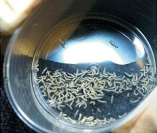
Sterile maggots from the common green bottle fly, Lucilia sericata, produce potent proteolytic enzymes and can consume up to 75mg of necrotic tissue per day as well as being capable of destroying bacteria11 (Figure 5). They are applied to the surface of a saline-cleaned wound and prevented from falling out by applying a woven mesh that allows them to receive oxygen to prevent them suffocating.
They are very useful in non-healing wounds or penetrating wounds of the hoof. Typically, 100 to 200 maggots are supplied and are relatively inexpensive (£150 for 100 maggots). Medical maggots are not available due to veterinary licensing restrictions; however, once this issue is resolved, they are a useful way of treating appropriate wounds.
Wounds result in undermining and loss of tissue, which creates dead space, reduces capillary perfusion and distracts the wound edges6. The elimination of dead space is fundamental to successful wound healing. Dead space can be eliminated by the use of drains, suturing and by packing the wounds with collagen-like biomaterials.
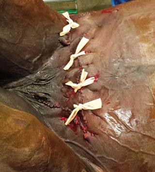
Drains are a useful way of eliminating dead space by preventing build-up of exudate or serum in the wound. Passive drains use gravity flow to wick or drain exudate down a non-adherent sterile material. Penrose drains are non-irritant and most commonly used. These are placed in a remote part of the wound and can be exited either through a separate incision adjacent to the wound or at the most distal aspect of the wound, which is left open (Figure 6).
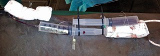
Vacuum-assisted wound closure may be beneficial for those unlikely to drain by gravity. It applies negative pressure to the wound, removing accumulated fluid – resulting in improved wound perfusion and decreases in wound infection rates12. While vacuum-assisted drains are commercially available, a cheap and effective way of creating one uses a 60ml luer lock syringe and an extension set placed into the cavity with a 14-gauge needle locking the plunger in a maximally drawn-up position (Figure 7).
These allow accurate measurements of accumulated fluid and removal at a point when drainage is known to be decreasing. Drains are sutured in place using a Chinese finger trap suture and can be fenestrated to minimise the risk of blocking. Drains will often block, but gentle irrigation with saline and suction is usually all that is required to remove the blockage. Drains can be left in until there is minimal discharge evident; however, after four to five days they may become a source of contamination and result in exudate and should be removed at this stage.
Every attempt should be made to suture both partial and full-thickness wounds unless there is too much tension, a risk of penetrating underlying synovial structures or significant tissue loss.
Restoration of the skin-to-skin contact with a bleeding edge maximises primary intention healing, thereby minimising time and scar formation associated with the healing process. The elasticity of the skin can be used to enable stretching across wounds in which significant retraction of the skin has occurred – and wounds associated with significant swelling can be cleansed, debrided and sutured once the swelling has reduced. Reduction of dead space using suture techniques has to be weighed up with the risk of creating a nidus for infection to persist.
Suture type depends on the location of the wound, the type of tissue being sutured, and the degree of tension under which the sutures are placed. Ideally, a monofilament absorbable or non-absorbable suture with high memory is used as this results in less tissue drag, is easier to suture, has less risk of inadvertent suture contamination with contaminated surfaces and less infection risk. The goal is to select a suture similar in strength to the tissue in which it is to be used13.

Blood flow to the skin edge is inversely proportional to the wound closure tension14 and sutures should be placed to minimise excessive tension at the skin edges. Loosely apposed skin edges are favourable; however, the reality is, in some wounds, significant tension is required to appose the skin edges15.
The use of tension-relieving suture patterns (near-far-far-near or horizontal/vertical mattress) at points of greatest tension, with interrupted sutures placed evenly distributed between them, can greatly facilitate wound closure, reduce tension on the suture and, therefore, the skin and allow apposition of skin edges that would cause breaking of a normal interrupted suture pattern.
The near-far-far-near suture pattern is optimal as it provides apposition of the skin edge in addition to providing tension relief. A larger suture diameter may be better if tension exists, although more sutures of smaller diameter are optimal rather than increasing suture size16. Small diameter sutures can be used between tension-relieving sutures. Undermining of the skin edges helps to preserve the blood supply as well as relieve tension.
In addition, the use of sterile Backhaus towel clamps (Figure 8), 1cm full thickness, tension-relieving incisions placed remotely from the wound, and small presterilised silicon quills used to incorporate the sutures either side of the wound, all help to relieve the tension at the wound edge and facilitate healing by primary intention (Figure 9).
Significant swelling and oedema of wound edges and adjacent regions will occur within hours of the injury. This can preclude suturing of the wound and, therefore, result in delayed closure. Application of a firmly placed bandage with a hypertonic saline dressing can help to reduce oedema of the tissues and more generalised swelling that will allow closure 24 to 48 hours post-injury.
As a rule, sutures should be placed at a distance from the skin edge equal to the thickness of the skin itself, but, invariably, tension of the wound and stiffness of the skin need to be considered15. Sutures should be placed a minimum of 0.5cm from the skin edge for maximal security and away from newly epithelialised tissue15.

Suture removal is usually performed between 10 and 14 days following wound closure. Wounds under tension will normally benefit from sutures being removed in stages to try to prevent dehiscence of the wound. It is important adequate protection of the wound remains in place following suture removal. Minimising movement of the limb and bandaging of the affected area is recommended for a minimum of a week following suture removal.
The composition of the wound changes as healing progresses; therefore, using appropriate dressings for the stage of healing will require changing the type of bandage applied to achieve optimal results17. The type of bandage material applied to the wound, therefore, should constantly be reassessed with each stage of healing to maximise the rapid progression of the particular healing stage. Bandages should supply protection to the wound, provide sufficient, evenly distributed pressure to the limb, and be applied so no movement of the bandage occurs.
Bandaging contributes to local hypoxia, which stimulates angiogenesis and the accumulation of exudates on the dressing against the wound surface, which provide a constant source of inflammatory mediators18. However, bandaging reduces contamination, protects vital structures, reduces oedema and provides mechanical stabilisation.
Primary layers are in direct contact with the wound and change depending on the stage of healing.
Secondary layers (for example, cotton roll, combine cotton) hold the primary layer in place and can provide additional protection against bacterial colonisation as well as providing support, absorbing fluid and even immobilising the limb. A layer of woven cotton applies pressure and protects the first two layers from contamination.
A tertiary layer of elasticated or adhesive bandages may be added to add stiffness and pressure – holding the bandage in place. This layer may also be used to immobilise the region if cast material is used. It is important to provide adequate relief of pressure over areas such as the accessory carpal bone by creating an opening in this region with a scalpel blade (Figure 10).
Irrespective of the wound, the creation of a moist wound environment to allow optimal healing is widely recognised across the human and veterinary fields19,20. The advantages of moist healing include:

Wound exudate in the absence of infection provides a substrate rich in enzymes, growth factors and chemotactic factors and provides the foundation for successful wound healing. With this in mind, the following principles can be applied when selecting appropriate dressings and topical products to apply to the wound.
Generally, for dry, untreated wounds, an amorphous hydrogel gel or bandage that contains glycerin, polymers and water is used to hydrate the wound. Once hydrated, these are discontinued in favour of more occlusive dressings.
Continued debridement of the wound requires a dressing material that removes necrotic tissue and bacteria. Honey and sugar-based bandage materials, as well as hypertonic saline dressings, can all be used during the debridement stage and cause minimal damage to healthy tissues. Hypertonic saline works by osmotic action to desiccate the necrotic tissue and bacteria in the wound, and has been shown to be particularly effective in debriding and reducing oedema associated with the wound bed.
The promotion of granulation tissue necessitates a moist wound environment. This can be created by the use of occlusive wound dressings. Occlusive wound dressings encourage rapid autolytic debridement with less necrotic tissue, a bacterial and waterproof barrier, a decrease in pain associated with dressing changes and decreased wound healing time. They are not associated with increased infection rates.
Occlusive dressings include amorphous gels, calcium alginate dressings and foam dressings. Calcium alginate dressings are soft, non-woven fabric pads, composed of sodium and calcium alginate, a derivative of seaweed. Calcium in the dressing interacts with sodium in the wound providing a wound exudate that stimulates myofibroblasts and epithelial cells21.
They are reserved for moderate to heavily draining wounds and can absorb up to 20 times their weight in exudate, so reducing the frequency of bandage changes. The dressing easily conforms to the wound, can be rolled up and put into crevices in granulation tissue and is easily and painlessly removed. These dressings prepare the wound bed by stimulating granulation tissue. In wounds that lack excess exudate, but still require granulation tissue stimulation, the alginates can be pre-moistened prior to application.
Exposed bone provides a challenging healing environment as, if the periosteal surface dries, it has a high propensity to sequestrate. Drilling 2mm holes through the cortical bone and into the medullary cavity can create bleeding tracts through which cells can migrate and help to granulate exposed bone. In addition, the use of hydrogels impregnated with acemannan reportedly stimulates healing over exposed bone by increasing angiogenic and fibrogenic growth factors over the wound22.
Once a reduction of exudate is achieved, and there is sufficient granulation tissue, foam dressings encourage epithelialisation and prevent exuberant granulation tissue from forming. As semi-occlusive dressings, they provide a moist wound environment by increasing the temperature of the wound by 1°C to 2°C 10.
Exuberant granulation tissue occurs as a result of an inefficient and protracted inflammatory response in horses, an imbalance in collagen homeostasis and an inefficient inflammatory response in the horse that makes it susceptible to infection wound expansion. It results in delays in wound contraction and inhibits its epithelialisation.
Exuberant granulation tissue is debrided prior to the application of a foam dressing, and this can be left for four to seven days. At this stage an antimicrobial-impregnated gauze dressing can also be beneficial in preventing bacterial colonisation. This contains biguanide or chlorhexidine digluconate – which suppress microbial growth – that is incorporated into fabric. It is particularly useful for wounds close to synovial structures.
Commercially available products are marketed for topical application to wounds in the equine veterinary market. While some have their place at different stages of the wound repair process, many fail to confer any additional advantage to create a moist wound environment and appropriate dressing application, and may inhibit this process.
Topical antiseptics are effective against a wide range of bacteria; however, they do not penetrate necrotic debris well and do not reduce bacterial populations deep in a wound bed. As already stated, saline in combination with gauze was more effective than silver sulfadiazine or povidone-iodine solution in reducing bacterial loads. Alcohols, aluminium salts, boric acid, chlorhexidine, hydrogen peroxide, hypochlorite, iodine, povidone-iodine and silver nitrate have more detrimental effects on wound healing than they do beneficial effects at reducing bacterial numbers.
Topical antimicrobial agents do provide efficacy against bacteria in the wound bed with minimal side effects on wound healing. Silver sulfadiazine and fluphenicol-based antimicrobial sprays have the advantage of not being used systemically, so reducing concerns about antibiotic resistance. Such topical sprays can be used following the debridement of the wound; however, their use, either instead of, or beneath, an appropriate dressing is of questionable benefit and may impede the beneficial effects of the dressing or autolytic debridement process. If used, the choice of antimicrobial spray should be based on the culture of the wound, which should be non-exudative and superficial.
Amorphous hydrogels are designed to provide moisture to a dry wound and are useful in creating a moist wound environment at the start of wound healing. They contain water, glycerin and polymers, conform to the wound, are non-drying and provide a bacterial barrier and, eventually, a moist wound environment. Being completely occlusive they provide the necessary environment for autolytic debridement, thermal regulation and white cell migration. Once the wound is moist, use of hydrogels can stop.
Hydrogels are also used as vehicle deliveries for other wound medications, such as silver sulfadiazine and metronidazole, and the use of hydrogels containing acemannan reportedly stimulate healing over exposed bone by releasing fibrogenic and angiogenic cytokines.
Vulketan Gel contains a potent serotonin receptor antagonist (ketanserin) and has been shown to result in two to five times more success in closure of second intention wounds by reducing infection and development of exuberant granulation tissue24. Therefore, it is best used in the inflammatory phase (early) and repair (later) phases of healing. In the study, treatment began on days six to nine and continued throughout the length of wound healing. Distal limb wounds prone to formation of exuberant granulation tissue benefit from Vulketan Gel.

In the debridement and inflammatory stages manuka honey from the nectar of the manuka bush (Leptospermum scoparium) in New Zealand has been shown to be effective at reducing bacterial colonisation, increased healing rates and achieving superior debridement to hydrogels25.
It has been demonstrated some commercially sourced edible honeys can be as effective as manuka honey and, therefore, may provide a cheaper source. However, in the same study there were 18/29 honeys in which bacteria was grown – thus medical-grade products should be used26. Honey comes incorporated into dressings as well as a topical treatment and can be applied during the debridement and granulation stage of wound healing.
Finally, platelet-rich plasma (PRP), routinely used as a treatment for tendon and ligament lesions, can also be applied to wounds with the addition of 10% calcium chloride to form a gel. Equally, PRP can be sprayed using a special applicator tip that mists the solution on to the wound. It provides a rich source of growth factors and, thereby, reduces exuberant granulation tissue and promotes wound contraction and epithelialisation, as well as inducing an anti-inflammatory response.
The common use of corticosteroid ointments and creams as a means of controlling exuberant granulation tissue has been shown to be contraindicated as they inhibit proteolytic matrix degradation and re-epithelisation27, both of which are essential for rapid wound repair. The moist wound environment and effective wound debridement is more effective and not detrimental (Figure 11).
It is important to ensure the horse is vaccinated against Clostridium tetani. If the tetanus status is unknown then administration of the tetanus antitoxin is essential. Antibiotic use should be used with discretion. In the initial stages of wound healing, antibiotics are recommended to prevent bacterial overload; however, once granulation tissue is forming, the use of antibiotics should be regularly reviewed. Effective antibiotic treatment should be orientated around culture and sensitivity; however, broad-spectrum antibiotics are probably necessary in most wounds given their degree of contamination.
Anti-inflammatories are used judiciously to control pain, minimise the inflammatory phase of healing and the formation of excessive granulation tissue. There are no deleterious effects of NSAIDs in wound healing28.
Some facts about wound healing in horses
Uniqueness of wound healing in ponies