20 Mar 2025
Medical imaging is a fundamental pillar of veterinary diagnostics, and is increasingly utilised in treating a variety of conditions in both human and veterinary medicine. According to Manuel Pinilla, recent advancements have significantly expanded the capabilities of imaging modalities, improving diagnostic accuracy, clinical efficiency and patient outcomes…
Manuel Pinilla
Job Title
Image: Adobe Firefly / AI generated
In veterinary medicine, access to advanced modalities has rapidly increased, both through referral centres and in first opinion practice.
Artificial intelligence (AI)presents further opportunities to improve image quality, data extraction and analysis, and diagnostic information. However, these advancements also present challenges, with a shortage of trained radiologists and radiographers and inconsistent quality control. This article explores the latest developments in veterinary diagnostic imaging, and how advancements in human imaging may translate into this field.
Ultrasound has become a mainstay of diagnostics in practice, although expertise varies greatly among users. In he past few years, protocols for regional, localised scans have become more commonplace, such as abdominal/thoracic-focused assessment with sonography for trauma (A/TFAST), and single organ scans. While faster and typically easier to learn and interpret, these approaches risk missing pathology from a more thorough cavity-screening procedure.
Peripatetic ultrasound services are expanding, allowing first opinion practices to access imaging expertise beyond the in-house team. These services also provide training opportunities, up-skilling staff to make better use of the practice machine and improve confidence. Remote services can also enable observation and training on live cases in the clinic through technological integrations.
Newer augmented reality for ultrasound enables interactive 3D visualisation, useful for surgical planning, client education and veterinary training. Use of 3D virtual reality echocardiography in human medicine provides enhanced cardiac assessments and is starting to enter the veterinary space.
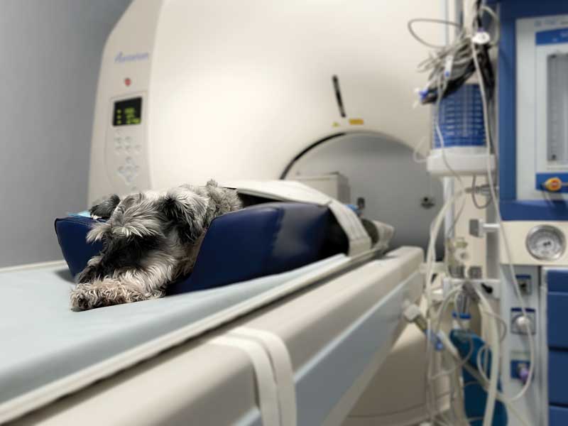
The past decade has seen a rapid increase in the installation of computed tomography (CT) machines in first opinion practice, increasing availability for a broader range of cases. Multidetector CT provides exceptional detail for most body areas, regardless of patient size. Cone beam CT is best adapted for dental and exotic animal imaging, providing enhanced spatial resolution compared to x-rays, typically at a lower cost than multidetector machines.
The growth of teleradiology services has increased access to specialist radiology reporting for advanced imaging. Radiologists also provide valuable input on appropriate modality selection for cases and accurate image acquisition. With the increasing use of remote services, training and support on case selection and machine use will ensure the right imaging is performed for the right cases, and/or including specialist input earlier in the diagnostic pathway (Pinilla, 2025).
The ability to create 3D reconstructions from CT scans has enabled the design of implants for precise surgical planning, particularly in orthopaedic and dental applications. Companies such as Perspectus Tech and Surgeree are combining this with virtual reality (VR) to enable users equipped with a VR headset to upload scans for viewing in 3D in a matter of minutes. This has tremendous utility in teaching anatomy and imaging, and opens up the potential to show clients the pathology of their pets in a novel, immersive way.
Positron emission tomography (PET) CT combines the anatomical information from CT with functional information from biomarkers. This integration allows for superior detection of tumours and metabolic activity, providing exceptional diagnostic capability.
Widely used in human medicine, developments include tracking CD45 expression to assess inflammation through imaging (Salehi et al, 2025). While cost has restricted its use in the UK and Europe, PET-CT is increasingly used in the veterinary field in both research and clinical practice in the US.
Radiomics is the extraction of quantitative information from medical images, including complex patterns that are difficult to recognise or quantify by the human eye (Mayerhoefer et al, 2020). It is a rapidly evolving field of research that captures tissue and lesion characteristics that may aid in the identification and classification of pathology. The use of radiomics in CT can improve the detection, staging and management of diffuse lung diseases in human patients. Latest studies have shown quantitative CT analysis may be useful in diagnosing and monitoring similar conditions such as bronchomalacia in dogs, documenting parenchymal changes that cannot be diagnosed with traditional thoracic radiography (Gerhard et al, 2024).
In future, radiomics may aid in predicting response to treatment and prognosis when applied to PET-CT scans and used in combination with other clinical data.
A comprehensive overview of AI in diagnostic imaging is beyond the scope of this article as it would very quickly become outdated in this fast-moving field of development. However, there are some broad principles of AI applications in imaging that warrant mentioning:
Enhancing efficiency: automated image labelling, orientation correction, and optimisation to speed the process of image interpretation.
Image quality improvement: AI processing to improve accuracy and reliability of images.
Diagnostic probabilities: AI “reading” of images to triage, highlight abnormalities and indicate the likelihood of pathologies. While few of these tools exist in veterinary radiology, this is a rapidly growing field. In human biomedical imaging, an entire library of tools exists to aid radiological interpretation.
Synthetic imaging: creating artificial lookalike images to increase database examples, for example generating variations of “normal” thoracic x-rays for teaching purposes.
Image-to-image translation: this is the conversion of images from one form to another, increasing accessibility to complex images, saving time and reducing costs. This can include conversion between different MRI sequences, followed by MRI to CT, and CT to other modalities (Zhong et al 2024).
Image mapping: AI can be used to map imaging findings from one species to another, or one modality to another. For example, research is ongoing to explore correlations between the MRI findings of cavalier King Charles spaniels with photographs of the head, with the hope of improving access to faster, less costly diagnosis of Chiari-like malformation and syringomyelia.
Standards and regulations: much discussion among global veterinary organisations and regulators is about ensuring appropriate safeguards are in place to ensure the reliability and accuracy of AI-assisted imaging.
Interventional radiology: one of the fastest areas of development in veterinary medicine has been the use of minimally invasive procedures using radiology, such as chemoembolisation and stent placement.
Contrast agents: with the potential to greatly enhance both anatomical and functional information from images, development of new contrast agents provides additional potential gains. For example, MRI contrast agents that are less potent than gadolinium may reduce the risk of side effects, and ultrasound contrast bubbles with a protein-copolymer coating for longer persistence improve ease of use and diagnostic utility (Tatiana et al).
3D ultrasound elastography: this technique measures tissue stiffness in response to an applied mechanical force (compression or shear wave). This allows assessment of changes in the elasticity of soft tissues resulting from specific pathological or physiological processes. For example, solid tumours often differ mechanically from surrounding healthy tissues and fibrosis typically increases tissue stiffness. In the human field, diffuse liver disease is the best validated application of elastography and has been adopted for non-invasive detection and staging of liver fibrosis and monitoring response to treatment (Sigrist et al, 2017).
Magnetic resonance spectroscopy (MRS): this technique allows non-invasive evaluation of tissue molecular composition, with metabolic and chemical analysis of tissues. The altered resonance of tissues where pathology is present can act like a “virtual biopsy”, reducing the need for more invasive biopsy procedures (Mariano et al, 2017). While largely restricted to research settings, increasing clinical applications are being explored, for example enhanced diagnosis, grading and follow-up in brain cancer patients (Weinberg et al, 2021).
Standing cross-sectional imaging machines: the development of CT and MRI units that enable limb scans in standing animals has revolutionised the accessibility and ease of imaging for the equine patient. Negating the need for general anaesthesia, the ability to obtain advanced images of the complex structures – particularly in the foot – without anaesthesia as an outpatient procedure has vastly improved lameness diagnosis for large animal patients.
4D imaging: the combination of 3D imaging with time allows movement and variation to be observed and analysed more accurately, and reduces motion artefact. This can be applied to ultrasound, CT, MRI and PET scans. Time-resolved 3D imaging is being applied in areas like radiation oncology and prenatal monitoring (Grace et al, 2021).
While many exciting developments in diagnostic imaging are on the horizon, important considerations are needed to ensure these technologies remain accessible and sustainable, benefiting patient outcomes. Even when innovations may offer significant advantages, their integration into veterinary practice is not without challenges.
Advanced imaging modalities require significant financial investment, which may not be feasible for veterinary practices and animal owners. The rapid evolution of imaging technologies has also outpaced the availability of trained veterinary radiologists and radiographers to acquire and interpret the images. Before conducting any diagnostic procedure – imaging or otherwise – veterinarians must also consider whether the results will provide meaningful insights that affect patient management. The ultimate goal is to improve patient outcomes by selecting the most impactful imaging modality. Where more choice exists, this can increase the complexity of clinical decision making.
The storage and transmission of high-resolution imaging data contributes to environmental impact. Solutions such as data compression will be necessary to balance increasing imaging demands with ecological concerns. AI applications also require the handling of large amounts of data. In addition, these technologies must address the challenges of the availability of representative, accurately labelled training data, data security and potential biases.
The field of diagnostic imaging is evolving rapidly, as technological advancements filter from human to veterinary medicine. Increased access to advanced modalities in both small and large animal practice is improving diagnostics, aiding more accurate clinical decision making.
AI-driven solutions, image mapping techniques and functional imaging have the potential to further improve diagnostic precision and patient outcomes. However, challenges such as access to expertise, the cost of advanced modalities, and data considerations must be addressed to ensure responsible implementation within the framework of contextual care. If these challenges can be met, the field of diagnostic imaging promises even greater accuracy, efficiency and accessibility, ultimately benefiting the health and welfare of more animals.