11 Nov 2019

Figure 1. A grade two open fracture of the radius and ulna in a dog. The dorsal aspect of the skin was penetrated by a spike of bone.
Open fractures are defined as any kind of fracture that communicates with the atmosphere or an unsterile region of the body (for example, nasal cavity, gastrointestinal, reproductive and urinary tract) through a wound of any size (Evans, 2016).
In small animals, open fractures are more commonly diagnosed on the distal limbs and the skull – particularly affecting the oral cavity, as there is less soft tissue coverage in these regions.
Epidemiologic studies have shown the incidence of open fractures in small animals is relatively low, usually being secondary to high-velocity trauma. Comminuted fracture configurations are reportedly more common (Millard and Weng, 2014).
In comparison with closed fractures, the management of open fractures is more challenging for the surgeon since the fracture, wound and interaction between these must be considered. In addition, soft tissue damage is often more extensive and the wounds are contaminated. These factors can lead to significant complications, such as delayed or non-union and infection, and ultimately can even result in amputation.
Emergency care for open fractures has to be performed promptly – the prognosis being adversely affected if the treatment is delayed.
The patient should be carefully assessed, and any life-threatening conditions should be prioritised before detailed inspection of the fracture. Open wounds should be covered with a sterile dressing to reduce the risk of nosocomial infections. A blood sample (cell count, biochemistry and electrolytes), ECG, abdominal ultrasound and thoracic radiography should be considered and, if facilities are available, oxygen saturation and blood pressure should be monitored.
Following the initial examination, oxygen therapy, analgesia, IV fluids and administration of blood products may be required. Once the patient is stabilised, the neurovascular integrity of the affected limb should be carefully evaluated. It is important to keep in mind some patients may respond negatively to nociceptive stimuli due to shock, therefore the neurological function of the affected limb may need to be re-assessed after 12 to 24 hours.
Analgesia should be considered as a priority in any patient following a traumatic episode. Opioids are used commonly; these can be combined with benzodiazepines if sedation is required. Administration of NSAIDs can also be considered, though these should be used with caution if any underlying gastrointestinal or renal disease is present, or if the patient is shocked and hypotensive.
Classification schemes for open fractures have been described taking into consideration the degree of soft tissue injury (Figures 1 to 3). These schemes can be useful as they can be used to predict whether a fracture is likely to become infected.
The key factors in prognosis of open fractures are the degree of soft tissue injury, the severity of vascular and neurological trauma, and the extent of contamination and/or infection.
The most widely used classification schemes have been suggested by Gustilo and Anderson (1976), and more recently, by the Orthopaedic Trauma Association (Evans et al, 2010; Panel 1).
Emergency wound treatment should be performed as soon as possible following cardiovascular stabilisation of the patient (Gustilo and Anderson, 1976; Patzakis and Wilkins, 1989). General anaesthesia or heavy sedation is likely to be required. After gross foreign material is removed, sterile gel should be applied to the wounds and the affected area clipped with a wide margin. The clipped area and the bordering skin can be prepped with 4% chlorhexidine gluconate solution or povidine iodine solution.
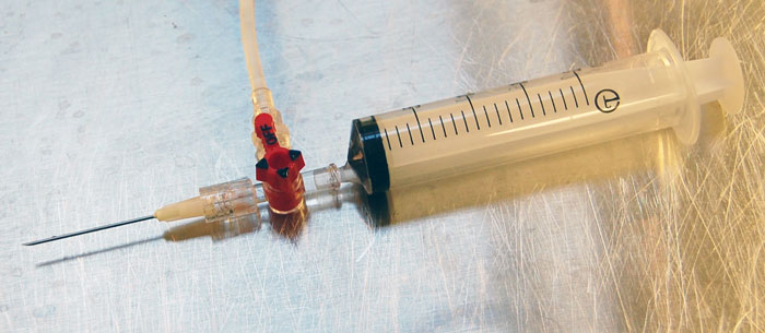
The wounds should be copiously lavaged with sterile isotonic fluids (sodium chloride 0.9% or Hartmann’s solution). The optimum pressure for wound irrigation is around 7psi to 8psi; this can be achieved using a 20ml syringe and a 19-gauge needle (Figure 4). It is helpful to cover the patient with an impervious drape during lavage of the wounds, to prevent unnecessary wetting and cooling of the patient.

Following initial irrigation, surgical layered debridement of the wound should be performed. This should be relatively conservative; if necessary, further debridement can be performed at a later date if more tissues become non-viable. Further irrigation following debridement should then be performed.
It is generally unhelpful to obtain swabs for bacteriology immediately post-debridement, as they are frequently unrepresentative of future infections (Lee, 1997). If signs of infection are subsequently encountered, swabs and aspirates from adjacent to the bone should be taken for culture and sensitivity.
Antibacterial therapy with a broad-spectrum antibiotic is typically initiated as early as possible post-injury (Patzakis and Wilkins, 1989). The most common bacteria found in acute wounds are Gram-positive – especially Staphylococcus species. Gram-negative bacteria – such as Klebsiella, Pseudomonas and Escherichia coli – are more common in chronic wounds and following bite wounds.
Appropriate antibiotics such as cefuroxime (20mg/kg IV every eight hours) or clavulanic acid-potentiated amoxicillin (20mg/kg IV every eight hours) are usually started immediately; oral antibiotics can be continued post-debridement, but these are generally discontinued after one to five days provided no further infection is suspected and good granulation tissue is present. If a concern regarding infection exists, continuation of antibiotic therapy should be based on culture and sensitivity.
The use of local antibiotics such as gentamycin impregnated collagen sponges, in conjunction with systemic antibacterials, can reduce and prevent the frequency of late or deep infections; however, these are thought to be unnecessary for the majority of patients.
Most grade one and two open fracture wounds can be closed primarily following debridement and irrigation, in conjunction with stabilisation of the fracture. If significant contamination is present or if fracture stabilisation is to be delayed, a period of open wound management can be helpful. Wet-to-dry dressings are ideal during this period.
Once granulation tissue has started to form, foam or non-adherent semi-absorbent dressings can be used. Wounds can then either be left to heal by secondary intention, or delayed primary or secondary closure can be performed at a later date.
For grade three open fractures with larger wounds and more contamination, a period of initial open wound management is usually recommended. In some cases, partial closure of wounds can also be considered; following suturing a small portion of a wound is left open to allow drainage.
Once any contamination has been controlled, options for definitive closure should be considered including second intention healing, delayed primary closure, secondary closure or use of reconstructive procedures using vascularised muscle and skin flaps or grafts.
It is important not to unnecessarily extend the period of open management as this can increase the infection rate and slow the bone healing; early use of vascularised flaps or grafts should be considered for larger defects. It should also be borne in mind that tissue stability is important for soft tissue healing, as well as fracture healing, and early stabilisation of the fracture can have significant beneficial effects on wound healing.
When choosing the appropriate method to stabilise an open fracture, the surgeon should consider a fixation method that provides adequate stability, but also maintains the integrity of the surrounding soft tissues. Fracture stabilisation should be performed as soon as possible following cardiovascular stabilisation of the patient; provision of fracture site stability is key for avoiding infection, as well as bone and soft tissue healing.
As type I open fractures are usually caused by low impact trauma, and the amount of soft tissue injury is minimal, these can essentially be treated as closed fractures. The AO principles should be applied, aiming to achieve a stable and robust fixation, and early return to limb function, therefore reducing the risk of complications (Panel 2).
Rigid fixation is achieved most reliably using internal fixation techniques such as bone plates and screws. Intramedullary devices such as the interlocking nail can also be considered; angle stable nails are particularly useful.
For simple fractures, absolute stability can be achieved by accurate reduction and compression of the fracture fragments; for comminuted fractures relative stability is achieved using bridging fixation, with no attempt to reconstruct intermediate fragments. Great caution is needed to minimise soft tissue damage at the fracture site, at the time of surgery; minimally invasive techniques can be used (Guiot and Déjardin, 2011).
Most type II fractures can also be managed effectively with a similar approach to closed fractures and type I fractures using internal fixation (Figure 5).
Open fractures may be slow to heal due to biological compromise; therefore, the implants need to be robust, as they may need to remain in place for an extended period of time. Combining implants – for example, use of a plate-rod or double plate construct – can be helpful, especially for comminuted fractures. Despite popular opinion, use of external skeletal fixators for the management of open fractures is often less optimal, since complication rates can be increased.
In addition, the healing times for open fractures may exceed the useful lifespan of an external skeletal fixator, necessitating revision surgery due to frame loosening.
In type III fractures, soft tissue damage is often more extensive. External skeletal fixation has traditionally been used to stabilise these fractures; however, since fracture healing is often slow, the risk of complications associated with external skeletal fixation is high, frequently necessitating revision surgery.
Therefore, internal fixation using bone plates and screws or an interlocking nail is often preferred. Use of internal fixation is generally relatively straightforward for fractures of the humerus or femur since the soft tissue envelop is extensive. Distal to the elbow or stifle internal fixation should be used where possible; however, it can be challenging to achieve soft tissue coverage of the implants in some cases.
Leaving implants such as bone plates exposed is to be avoided. In these cases, use of external skeletal fixation can be considered to provide initial stability and allow wound management. Once a wound has been closed or is granulating, a second procedure can be considered to remove the external skeletal fixation and apply definitive internal fixation. Alternatively, once contamination is controlled immediate internal fixation can be applied in conjunction with vascularised muscle and skin grafts.
Because healing of many open fractures may be delayed, cancellous bone grafting should be considered. Bone grafts can be applied at the time of initial stabilisation, as long enough soft tissue coverage is present; if soft tissue coverage is poor a delayed grafting procedure can be considered after a few days or weeks, once the wound has been closed.
As in any routine orthopaedic surgery, orthogonal postoperative projections should be obtained. Fragment apposition, limb alignment and implant positioning should be assessed. A soft bandage can be applied to the distal limb to reduce soft tissue swelling and to protect the wounds.
The bandage would typically be maintained for two to four days postoperatively, but this period may be extended to support contact dressings if open wound management is being performed. Appropriate analgesia is recommended and exercise should be restricted, although controlled exercise to allow gentle use of the limb during the healing period should be encouraged.
As aforementioned, antibiotics are usually discontinued after one to five days unless any infection develops. Follow-up radiographs will be obtained after around four to five weeks for paediatric patients and around eight weeks for adult patients. Radiographs are typically repeated every four weeks after this until fracture healing is documented, at which point unrestricted exercise can be resumed.
In most cases, implant removal is not required; however, owners should be warned of the possibility that a late occult implant infection could develop, in which case implants may need to be removed at a later date.
The main factors influencing prognosis in patients with open fractures include the extent of soft tissue damage and the degree of contamination. However, with appropriate emergency and definitive treatment the prognosis is surprisingly good – especially for grade one and two fractures.
Provision of appropriate fracture site stability using internal fixation is thought to give better results with fewer complications compared to traditional use of external skeletal fixators.