
03 August 2026
Lameness is a common issue in young dogs, but many owners will expect imaging from the start. This article looks at how to help clients see the options.

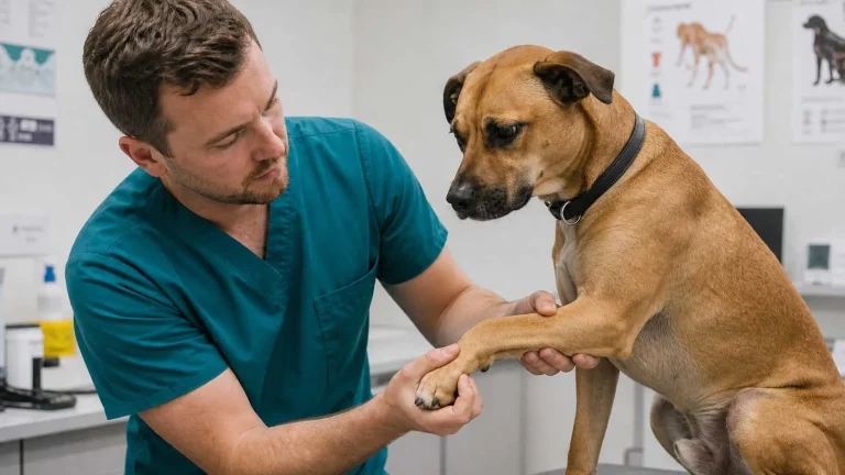
Lameness in young dogs is a common presentation, and it’s one where owners often expect (and ask for) imaging immediately. But as clinicians, you know that not every limp requires radiographs. Sometimes rest and observation are appropriate; sometimes targeted diagnostics are essential. The challenge is explaining this reasoning clearly, particularly when owners are anxious and want “everything done” to find out what’s wrong. This article explores how to navigate that conversation with confidence and clarity.
Charlie presents with a three-day history of mild left hindlimb lameness. The owner noticed Charlie was reluctant to jump up on the sofa the preceding day, but Charlie still plays and is eating normally. On examination, Charlie has grade 2/5 lameness on the left hind leg. Joint flexion and extension are normal, there’s no obvious swelling, and Charlie doesn’t show pain on palpation. He is neurologically normal.
Your initial impression: likely muscle strain, possible mild cruciate ligament strain or minor meniscal involvement. But nothing acutely obvious on physical exam.
The owner’s question: “Should we x-ray to see what’s wrong? I want to make sure it’s nothing serious.”
Recommend 1–2 weeks of restricted activity: lead-only exercise, no jumping or rough play, soft bedding and potentially short-term anti-inflammatory medication (NSAIDs). Recheck in 7 to 10 days.
Rationale: many cases of mild lameness in young dogs are self-limited. Muscle strains and minor ligament injuries often improve significantly with rest. If Charlie improves substantially by the recheck, imaging wasn’t necessary. If lameness persists or worsens, imaging at that point is more targeted and potentially more informative (you’ll have a clearer sense of what’s happening).
Cost now: £60 to £100 for a consultation and medication. Recheck in 10 days: £40 to £60.
Proceed with radiographs now. This provides immediate answers and rules out structural problems.
Rationale: if underlying pathology is present – such as early hip dysplasia, a small fracture or meniscal pathology – imaging will reveal it. This gives the owner reassurance and a clear plan.
Cost now: £100 to £150 for radiographs (depending on views). If pathology is found, additional consultation and treatment costs follow.
The evidence supports a stepwise approach: clinical assessment, conservative management and reassessment. But owners often want certainty now, and imaging provides that. The question is whether the cost and radiation exposure are justified given the clinical presentation.
For Charlie, with a short history, mild lameness and a normal physical exam, conservative management is evidence-based. But the owner’s desire for imaging isn’t unreasonable – it’s driven by anxiety and a desire to help their dog. Your job is to explain the clinical thinking clearly and help them understand that choosing observation first isn’t a compromise.
“Charlie is definitely lame on the left hind leg, and I want to help figure out why. What I found on my examination is that Charlie’s joints are moving normally, there’s no obvious swelling and Charlie didn’t react with pain when I felt around. That’s actually reassuring – it tells me we’re probably not dealing with something that needs emergency surgery.”
This is important: you’re not claiming to know the diagnosis. You’re saying what your examination ruled out or made less likely. This is honest and helps the owner understand your thinking.
“Given Charlie’s age and what I’m seeing, the most likely explanation is a muscle strain or a mild strain to one of the ligaments in the knee. These injuries often improve significantly with rest – similar to a sprain in humans. Charlie might benefit from limited activity for a week or two, and potentially some anti-inflammatory medication to help with any discomfort.”
This is the critical conversation. Many owners expect imaging; your job is to explain why it’s not the first step.
“Imaging – x-rays or ultrasound – is a useful tool, but here’s the thing: if Charlie has a simple muscle strain, imaging will show that the bones look normal, but it won’t show us the soft tissue damage. So we get information, but it might not change the treatment plan. Meanwhile, Charlie has been sedated and exposed to radiation. On the other hand, if we give Charlie a few days of rest and pain relief and see how he responds, we’ll learn a lot. If he’s better, we know it was likely a strain and we can avoid imaging altogether. If he’s not better, we can take x-rays then with a clearer idea of what we’re looking for.”
Owners need to know exactly what they should be watching for.
“Here’s what we’re looking for over the next week: is Charlie using the leg more? Can he jump up without pain? Is the limp improving day by day? If yes to those, we’re on the right track. But if Charlie is limping the same way a week from now, or if he’s not putting any weight on the leg, or if the swelling gets worse, those things would tell us we need imaging to dig deeper. We’ll recheck Charlie in 10 days, and based on progress, we’ll decide what comes next.”
Some owners will insist. That’s okay. Be flexible, but be honest.
“I understand you’d feel better knowing what’s going on. We can certainly take radiographs. Just know that if everything looks normal, the plan will still be rest and medication – the same as we’d recommend without imaging. But if it gives you peace of mind, let’s do it. And if we see something unexpected, that helps us plan next steps.”
“The cost of waiting and seeing is lower than imaging upfront. We’re looking at £60 to £100 now for medication and a consultation, then another check-up in 10 days. That’s about £100 to £150 total. If we did radiographs now, that’s another £100 to £150 upfront. Many insurance policies cover diagnostic imaging, so if you have insurance, it might be worth checking what’s covered.”
If you suspect cranial cruciate ligament disease based on clinical signs – positive drawer sign, tibial thrust or a history of lameness that worsens with activity – imaging is more clearly indicated. Dogs with likely cruciate disease benefit from early intervention and may develop secondary problems (meniscal tears, osteoarthritis) if left untreated. Be explicit if you’re concerned about this:
“Charlie’s signs are subtle right now, but if I was more convinced this was a cruciate issue, I’d be recommending imaging today. Since I’m not, we’re taking a stepwise approach. But if the lameness doesn’t improve or if specific signs develop, imaging becomes more urgent.”
Visit the Agria Cost of Care Conversations hub for more case-based guides