11 Apr 2016
Samantha Taylor
Job Title
Figure 1c. A visible needle piercing the palate. Image: © Melanie Spencer Marshall.
The vast majority of vets will have removed foreign bodies from the intestines of dogs, but perhaps credit cats with more selective tastes.
However, International Cat Care (ICC) carried out a small survey of vets as part of its Keeping Cats Safe campaign – for which it teamed up with The Veterinary Poisons Information Service and Agria Pet Insurance – and found all but one of them had treated cats with gastrointestinal foreign bodies.
This article will look at objects most commonly reported to effectively advise clients on what to keep out of reach of their curious cats.
Linear foreign bodies tend to be considered more common in cats than discrete objects and this was echoed in ICC’s survey; however, a study from first opinion practice showed only 33% of cases in cats were linear (Hayes, 2009).
Certainly, linear foreign bodies can carry a less favourable prognosis as, more frequently, they require multiple intestinal incisions for removal (Hayes, 2009; Hobday et al, 2014).
So, what did vets report as the most common intestinal foreign bodies seen in cats? Panel 1 lists some of the foreign bodies reported by veterinarians who took part in the survey.
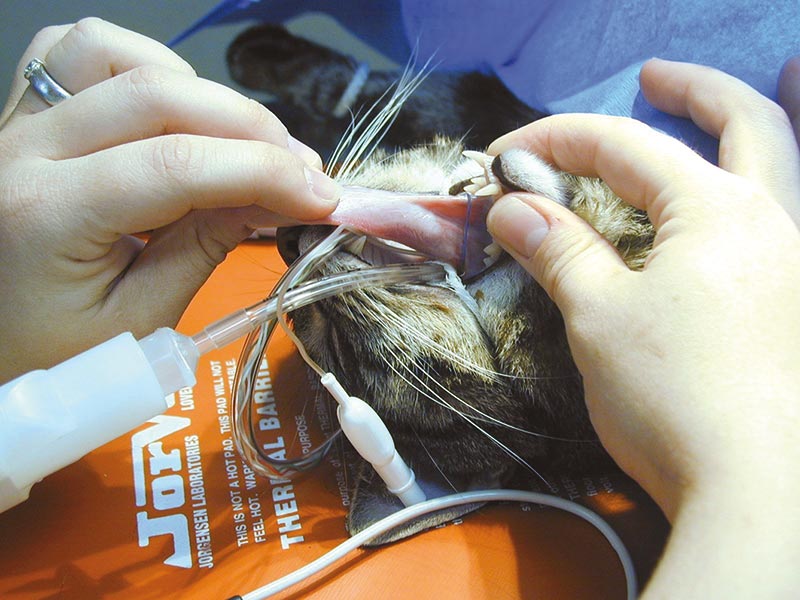
Needles and threads feature in all case series of foreign bodies in cats and ICC’s survey confirmed these as commonly reported in cats. Needle ingestion can result in dramatic presentations, such as penetration of the globe (Delgado, 2015) or even the brainstem (Cottam and Gannon, 2015).
More commonly, the needle ends up in the intestines along with the thread (Figure 1a), but can become lodged in the mouth (Figures 1b, 1c and 1d).
Hairbands and rubber bands were frequently mentioned, along with the string found around meat. Fibres and stuffing from inside cat toys, carpet fibres, ribbon, dental floss and blind cords all had a mention. Even the hair from an owner’s wig resulted in a linear foreign body in one cat, and Figures 2a and 2b reveal the impressive intestinal plication that resulted.
Small, round objects are just the right size to cause complete intestinal obstruction and seem irresistible to some cats. Coins (Figure 3) are not uncommon, together with buttons, earplugs, fruit stones, nut shells, bottle tops and almonds. One cat managed to swallow a pin badge, which caused a perfect intestinal obstruction (Figures 4a and 4b), necessitating an enterectomy for removal.
Many vets will have a story to tell about unusual objects removed from cats, and vets gave an impressive list, which included a beheaded plastic lizard (Figure 5), other children’s toys (Figure 6), an amazing radiograph of a cat that had eaten a small, spiky rubber toy (Figure 7), Christmas decorations and even a mobile phone SIM card.
Bones can also cause a problem to bin-raiding cats (Figure 8), as cooked chicken bones, in particular, can be very sharp and challenging to remove.
The vast majority of respondents in the survey described young cats being more likely to suffer from foreign bodies. Purebreds were frequently reported, with Burmese over-represented. Siamese and Siamese-related cats have been reported to suffer more commonly from pica – particularly, wool eating – and thus perhaps are at increased risk of linear foreign body ingestion.
Although not reported in published studies, ICC’s research suggests indoor cats may be more likely to ingest foreign bodies – emphasising the need for client education on environmental enrichment for cats kept solely indoors. Resources such as Ellis et al (2013) provide further information.
Clinical signs resulting from partial or complete intestinal obstruction vary depending on the location of the foreign body, which, in cats, tends to be the proximal gastrointestinal tract (Hayes, 2009). Vomiting, anorexia and, less commonly, diarrhoea are reported.
In cats, clinical signs of abdominal pain may be subtle, with lethargy and inappetence noted by owners. Cats may be presented after observed foreign body consumption, prior to development of clinical signs.
Intestinal foreign bodies may be expected to present acutely, but intermittent partial obstruction with clinical signs of more than a month’s duration is reported (Willis and Farrow, 1991), and the duration of clinical signs in Hayes (2009) was up to 30 days.
Cats with complete obstruction are likely to present with varying degrees of fluid volume deficits and severe cases with intestinal perforation will show signs of peritonitis and sepsis. Affected cats may show pain on abdominal palpation. The foreign body (or intestinal abnormality) may be palpated in up to 58% of cases (Hayes, 2009). Importantly, linear foreign bodies may be visualised anchored under the tongue (Figure 9) and any cat with consistent presenting signs should have the base of the tongue inspected.
Diagnosis may be based on history of foreign body ingestion and clinical signs, or the finding of a linear foreign body tethered at the base of the tongue. Imaging can be helpful in making a diagnosis of an intestinal foreign body. In this article, the author has shown some dramatic and obvious radiographs, but many cases are more subtle, with partial obstruction not always an easy diagnosis.
Non-radiopaque foreign bodies can be suspected in cases with dilation of the intestine proximal to the obstruction with gas and fluid accumulation. A study examining this diagnosis showed if the jejunal diameter is greater than two-and-a-half times the length of the cranial end plate of L2 then intestinal obstruction is the most likely diagnosis (Adams et al, 2010). Partial obstructions may not always result in dilation of proximal intestine at the time of imaging, however, making this a challenging diagnosis sometimes only made at surgery.
Linear foreign bodies causing intestinal plication may be suspected radiographically when the jejunum seems gathered and most of the small intestine appears bunched in one area of the abdomen, leaving a space on the lateral view. On the dorsoventral view the intestine may appear bunched on the right side.
Contrast radiography may be useful, especially in cases of partial obstruction, but should be used with caution if intestinal perforation is suspected.
Ultrasound has been shown to be very useful in diagnosing foreign bodies (Tyrell and Beck, 2006), although more challenging in cases with linear foreign bodies when plicated intestine or a hyperechoic line may be noted.
Prompt presentation and treatment is desirable. In the Hayes study (2009), cats that did not survive all had clinical signs of more than 14 days’ duration. Cats are masters at hiding illness and owners may be reluctant to bring their cat to the surgery due to perceived stress – preventing early diagnosis and, in the case of foreign bodies, significantly worsening the prognosis. Client education on watching for signs of illness, as well as running a cat friendly clinic (www.catfriendlyclinic.org) to reduce stress, may, therefore, help improve survival.
Treatment frequently includes surgical or endoscopic removal of the foreign body alongside management of fluid and electrolyte imbalances.
Conservative management is occasionally attempted – usually in specific cases, such as a cat with a linear foreign body tethered around the tongue. The thread is cut and the cat should be hospitalised and monitored closely with the owners counselled and practitioners prepared to go to surgery should the cat show signs of intestinal obstruction or perforation. The authors of one study (Basher and Fowler, 1987) treated 19 cats in this way, and 10 went on to require surgery.
Surgical enterotomy to remove a foreign body should be performed once a cat is stable and fluid and electrolyte abnormalities have been corrected. Discrete foreign bodies should be removed via an incision distal to the obstruction to ensure the healthy gut is sutured. Linear foreign bodies may require multiple enterotomies to remove them – to avoid excessive traction and resultant intestinal damage, following release of any anchor points (tongue or pylorus, most frequently).
Foreign body ingestion remains a differential diagnosis for gastrointestinal signs in cats, with purebreds, indoor-only cats and young cats over-represented in ICC’s small survey. Cats with partial obstructions may present with a more chronic and subtle history.
Owners should be advised on the nature of potential foreign bodies and be encouraged to store objects, such as needles and threads, securely. Indoor cats should be provided with safe, stimulating toys and their environment examined for any deficiencies.