25 Mar 2025
Gabriele Ghirello
Job Title
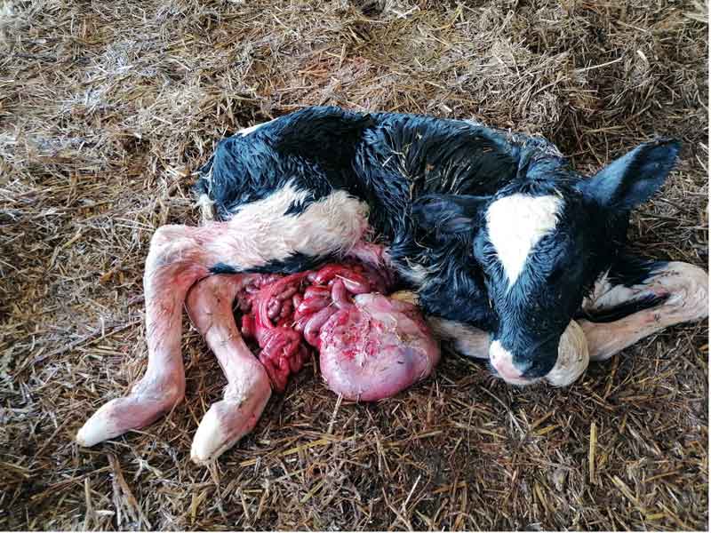
Figure 1. Fetal anasarca. This condition, common in dogs, cattle and sheep, can be of immune or non-immune causes and is a common presentation of aborted fetuses, often leading to complicated delivery.
Abortions can be an economic and emotional challenge in livestock farming. While a certain number of unsuccessful pregnancies is unavoidable, an increase in number and/or concentration of abortions might suggest husbandry or health risks on farm. Abortion rates of 2% to 3% are normal, while higher rates should be investigated by a veterinarian.
An abortion is the interruption of pregnancy after the organogenesis of the fetus is complete and before the fetus is viable. In cattle, this corresponds to days 45 to 265 of gestation, while in ewes and goats, this corresponds to days 45 to 140 of pregnancy.
Prior to this, pregnancy losses are considered embryonic deaths, and the embryo is often reabsorbed or mummified. These are usually difficult to identify, unless returns to heat are adequately detected.
Diagnosing the cause of abortions can be challenging. The fetus is often expelled days after its death, or weeks after the initial infection, therefore it can be difficult to isolate the pathogen responsible for the fetal loss.
Abortions can be caused by infectious/parasitic and non-infectious causes.
Bluetongue is a notifiable, non-zoonotic, viral disease. It is spread by midges belonging to the genus Culicoides. Although usually present in warmer areas, it is currently spreading concern to northern Europe due to the ability of midges to travel long distances. The first cases of bluetongue in the UK were identified through surveillance in November 2023.
Twenty-nine serotypes of bluetongue virus (BTV) have been identified worldwide. Sheep are affected most severely; however, cattle, goat and camelids can also be affected. Affected sheep present with fever, lethargy, facial swellings, nose and mouth erosions, sialorrhea and nasal discharge. Although less frequent and usually less severe, similar signs can be found in cattle. Transplacental transmission of BTV has been documented and confirmed to cause abortions and fetal malformations.
Diagnosis is based mainly on clinical signs, virus detection via PCR and serology. Treatment includes supportive care and antibiosis for secondary infections.
Control of bluetongue is mainly through vaccination and control of midges’ populations. However, currently vaccination does not generate cross-immunity between serotypes.
Bovine viral diarrhoea (BVD) is a complex viral disease of cattle (caused by a Pestivirus) that can cause abortions and major financial losses in UK herds. It is spread via contact, and sexual or transplacental transmission. The conceptus, infected in utero, can reabsorb (if infected within 40 days of pregnancy), become a persistently infected (PI) calf (between 40 and 130 days) or lead to abortion or congenital defects, when infected after 125 days.
Diagnosis is performed via blood (ELISA antibodies or antigen), milk or tissue samples (in vivo for screening or on aborted material). Prevention consists of early identification and removal of PI calves and vaccination.
Similar to BVD in cattle, border disease (BD) in sheep is caused by another Pestivirus. In naive flocks, infection can lead to abortion or mummification in any stage of the pregnancy.
In sustained pregnancies, affected lambs (“hairy shakers”) are born weak or with neurological signs. An increase in barren rates within the flock and birth of underweight and neurological lambs are suggestive of BD. Diagnosis is done via histopathology of aborted material, blood serology or tissue PCR. Currently, no effective vaccination or treatment exists. Removal of PI animals and a closed flock policy are the best way to control the disease.
Brucellosis is a zoonotic, notifiable disease of cattle (Brucella abortus), sheep (B melitensis and B ovis) and goats (B melitensis), last seen in cattle in the UK in 2004. In cattle, it causes abortion in the second half of gestation, or premature calves. In ovines, it causes abortion in ewes and orchitis in rams.
Transmission is mainly vertical (in utero) or through direct contact with aborted material or infected bodily fluids. Aborted material can be tested for diagnosis, alongside dam’s serology.
Campylobacteriosis is a zoonotic disease caused by Campylobacter, leading to embryonic deaths and abortions in the second half of gestation in cattle and sheep. Transmission is either venereal or through ingestion. No vaccines are currently available in the UK for cattle or sheep, but can be sourced via import.
Control is mainly performed through closed herd/flock policies, use of AI and farm hygiene during outbreaks. Diagnosis is achieved through bacterial culture of placental and fetal material.
Chlamidophila abortus is causative of enzootic abortion in ewes and goats and can cause sporadic abortion in cattle. Infection can happen at any stage of gestation, but usually causes late-term abortions, namely within the final three weeks of gestation in does and ewes and in the final trimester in cattle.
Animals are immune after abortion. Fetuses are often fresh, and necrotic placentitis is often observed. Diagnosis is through ELISA, PCR, antibody staining or isolation.
The most effective way to reduce major economic losses due to enzootic abortion in sheep and goats is vaccination prior to mating. There are licensed products for sheep, but no vaccination available for cattle.
C abortus is zoonotic and abortive in women also.
Infectious bovine rhinotracheitis (IBR) is caused by bovine herpesvirus-1. It is highly contagious and widely spread across the UK. It causes both respiratory (upper tract infections) and reproductive (pustular vulvovaginitis) diseases in cattle, which can lead to sporadic abortions.
Infection is via direct contact with infected animals, contamination through bodily fluids and aerosols, or vertical through the placenta. Abortion can happen at any stage of gestation, but happens more often in the second half.
Diagnosis is via immunologic staining on aborted material and placenta. In dams, serology or PCR can help differentiate between latent and active infections. Control is performed through routine bulk tank screening, biosecurity, removal of sources of infection and vaccination.
Several protocols are available, including modified-live vaccines and killed vaccines.
Leptospirosis can cause abortion in cattle and goats. Sheep are usually resistant to it, but can carry and excrete it. Therefore, mixed grazing increases the risk for cattle. It is more common in grazing flocks during pasture months, especially around watercourses. Leptospira borgpetersenii serovar Hardjo and Leptospira interrogans serovar Hardjo are among the most common in cattle in the UK and cause abortion in the third trimester.
Dams can abort or have premature, weak calves without being clinically affected, or show signs of fever, sudden milk drop, jaundice, anaemia and placentitis. Aborted fetuses and placenta can be submitted for antibody staining or PCR.
Adult females can be tested via PCR and serology or screened via individual milk or bulk tank test (non-vaccinated herds). Multivalent vaccines are available for the control of leptospirosis in cattle, and sources of infection should be identified as part of the control.
Leptospirosis is a zoonotic disease.
Neosporosis is a common parasitic disease that can cause abortion in cattle, sheep and goats. It is caused by protozoa Neospora caninum. Canids are definitive hosts and shed oocysts after eating tissues of infected animals, and intermediate hosts, such as cattle, become infected by ingesting oocysts. Transplacental infection to the fetus may then occur and cause abortion.
Abortions usually happen mid to late gestation, from dams that are often clinically unaffected. Fetuses are otherwise often born premature, as stillbirths or with cardiac and neurological complications.
Diagnosis is usually performed with PCR detection and immunohistochemistry on fetal tissues and serology on the dam.
Control of neosporosis involves limiting contact of farm and public dogs with cattle, reducing contamination of feedstuff and water sources, limiting the dogs’ ingestion of aborted material and dead stock and replacing animals from seropositive dams.
Q fever is caused by the bacterium Coxiella burnetii, which causes abortions in ruminants. A 2017 AHDB study has revealed the presence of antibodies for Coxiella in 79.8% of the bulk milk samples, making it a widespread disease across the UK. It is a zoonosis and, therefore, also a cause of abortion in humans.
It is mainly spread via bodily fluids, such as faeces, milk, urine and fetal membranes and fluids, with infection occurring via inhalation or contact with the bacteria. Due to a high concentration in amniotic fluid, it can put those assisting births at risk.
Common clinical signs of the infection include late abortions, stillbirths, higher incidence of metritis, endometritis and retained fetal membranes. However, most of the time, the infection is subclinical, and is, therefore, more challenging to identify.
Diagnosis is done via PCR testing on bulk milk or via serology.
The use of antibiotics to control Q fever has limited efficacy. In cattle, evidence shows the efficiency of antibiotic treatment at drying off in reducing shedding at calving, but not of reducing the bacterial load in infected animals.
In sheep, antibiotic use has not been proved to reduce the bacterial load.
Prevention includes hygiene during and after birth, with prompt removal of suspected aborted materials. Vaccination is available for cattle, sheep and goats, to control clinical signs of the disease, prevent abortion and to reduce shedding.
Salmonella infections can cause abortion storms in cattle and small ruminants, with or without systemic and enteric disease. Transmission occurs via contact with contaminated facilities, ingestion of contaminated feedstuff, aborted material and milk. S Dublin and S typhimurium are the most common serotypes causing abortion in cattle and sheep in the UK, alongside S abortusovis in sheep.
Abortions occur in the second half of pregnancy. Premature births and stillbirths can also happen. Treatment of infected animals involves antibiosis and supportive care.
Prevention involves isolation of affected animals, disinfection of facilities, vermin and bird-proofing feed stores, and vaccinating herds with a history of salmonellosis.
The Schmallenberg virus, spread by Culicoides midges, is often associated with fetal malformations, stillbirths and abortions in ruminants if infection occurs between two and six months of gestation in cattle, and between the first and second months in sheep. Acute infection of dams often results in pyrexia, scours and reduced milk yield in cattle, while this is not often observed in sheep.
Diagnosis is through PCR testing of fetal brain and serology of the dam.
Control involves reducing the exposure to the midges and planning the mating period accordingly.
Toxoplasmosis is a zoonotic parasitic disease of sheep and goats caused by Toxoplasma gondii. It is a major cause of fetal reabsorption within the first two months of gestation, and abortion and mummification when contracted later. The disease is transmitted through ingestion of oocysts from feedstuff or pasture contaminated with cat faeces. After infection, sheep become immune.
Diagnosis is confirmed through antibody testing and histopathology on fetal tissues. Control of the disease entails rodent control, stable farm cat populations, vaccination ahead of tupping and keeping on-farm animals previously exposed to the disease, which are therefore immune for life.
Several other pathogens potentially cause abortion in ruminants. Theoretically, any infection ascending from the vagina could put the viability of the fetus at risk, if causing a severe enough inflammation of the placenta or compromising the animal systemically.
Other pathogens that often cause abortions are:
Abortions of fungal aetiology are common, often due to Aspergillus species. These are often introduced via the oral or respiratory route and originate from mouldy or poorly prepared feed. Mycotic abortions usually happen in the second half of the pregnancy and are usually the consequence of a severe placentitis.
Trauma or stress can cause abortions. Transport, movement of animals or fights can cause fetal loss at any time of gestation.
Heat stress, from environmental temperatures or fever, and leading to fetal hyperthermia, can also contribute to fetal losses.
Nutritional causes, such as hyper and hypo-vitaminosis (vitamin A or E) or mineral imbalances (selenium, iron or iodine) can affect the development of the conceptus and cause abortion. Exposure to toxic plants, phytotoxins or mycotoxins can all cause fetal losses.
Therapeutics commonly used in farm practice are abortifacient. These include prostaglandins, corticosteroids (for example, dexamethasone) and oxytocin, when used inappropriately.
The management and prevention of abortion outbreaks can be challenging for farmers and veterinarians. A successful approach includes: