20 May 2019
Sonya Miles
Job Title
Rats are affectionate, interactive and popular pets (Tamura, 2010), and one of the most commonly presented exotic mammals in veterinary practice.
Soft tissue masses, as well as respiratory disease, are the main reasons pet rats are presented to vets. For the purpose of this article, the author will concentrate on mammary masses.
Rodent mammary tissue is extensively distributed in both sexes, with tumours occurring anywhere from the neck to the inguinal region (Hillyer and Quesenberry, 1997). The most common type of mammary mass is mammary gland fibroadenoma (Capello, 2011).
A good prognosis of survival often follows complete surgical removal of benign fibroadenomas in pet rats (Hillyer and Quesenberry, 1997; Capello, 2011), as they rarely metastasise (Capello, 2011); however, histopathological analysis is always recommended to confirm tumour type (Hillyer and Quesenberry, 1997).
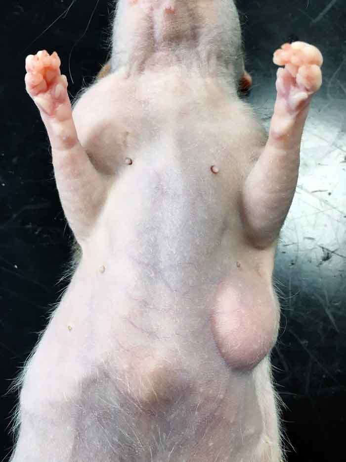
A two-year-old entire female rat – weighing 0.28kg (body condition score three of five) – was presented with two, approximately 2.5cm mobile masses on the right side of its neck and left axilla (Figure 1).
The masses had been visible for seven days, but due to the skittish nature of the rat – and lack of handling from the owner – may have been there for longer. The rat’s housing and diet were suitable for her species.
On initial examination, thoracic auscultation and abdominal palpation were normal. No nasal or ocular discharges were noted. The masses were found to be firm, mobile and non-painful. No other masses were found.
Due to the inability of rodents to vomit, no preanaesthetic fasting was required (Longley, 2008) – therefore also minimising the risk of hypoglycaemia, which is of concern in rats due to their small size (Capello, 2011).
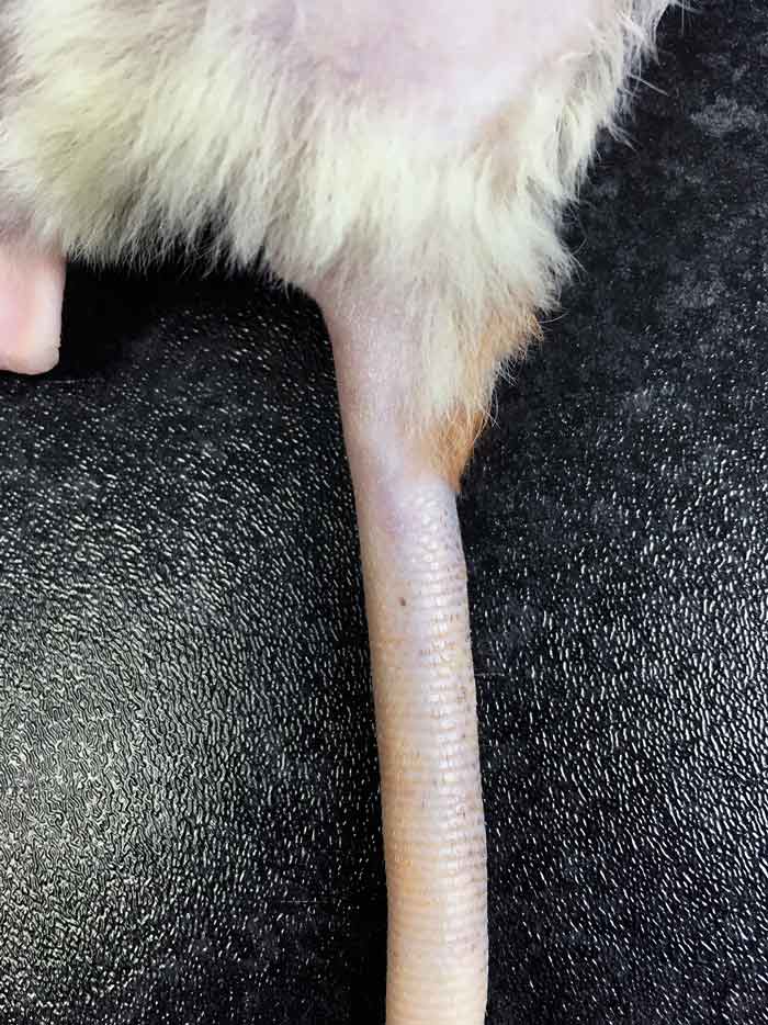
IV access was gained, using one of the lateral tail veins (Figure 2) – allowing the rat to be given 5ml of sterile Hartmann’s solution during surgery.
The rat was given a premedication of 0.05mg/kg buprenorphine IV. Half an hour later, it was placed in an induction chamber and preoxygenation was performed for five minutes, before it was anaesthetised with eight per cent sevoflurane 3L/minute of oxygen.
Once the rat had lost its righting reflex, a Darvall breathing circuit was used to allow sevoflurane maintenance at four per cent. The rat’s mouth was checked and cleared of food to minimise the risk of aspiration.
The rat was placed into lateral recumbency, and the surgical site clipped and aseptically prepared using diluted chlorhexidine. Using a concentrated version of the same preparation, the vet surgically scrubbed to aid asepsis. Eye lubrication was applied to the rat to prevent corneal ulceration.
As hypothermia is common in anaesthetised rodents (Longley, 2008), the patient was placed on a warming system and its extremities were wrapped with bubble wrap. Rectal temperature was monitored throughout to ensure it was maintained within its optimum range. Heart rate and rhythm were monitored using Doppler ultrasonography over the apex beat, while clear, sterile drapes were used so respiratory rate and effort could be monitored throughout.
Using a number 15 blade, an incision was made over the mass. Electrosurgery was used to cauterise small bleeding vessels, preserving blood volume. The masses were located, and blunt dissected away from the surrounding SC tissue and underlying musculature. Large blood vessels at the base of each mass were ligated using 4/0 poliglecaprone, allowing both entire masses and surrounding mammary tissue to be removed.
The surgical sites were instilled with dilute lidocaine not exceeding 4mg/kg (Carpenter, 2005) prior to the SC tissues being closed with a simple continuous pattern using 4/0 poliglecaprone. The same suture material was then used to close the skin using an intradermal pattern, employing an Aberdeen knot to close (Stott et al, 2007). This suture material was chosen for its monofilament and absorptive qualities, which, in this instance, would allow ideal wound healing and repair. In this case, an ovariectomy was declined by the client, despite being recommended.
Once the rat was recovering from anaesthesia, meloxicam at 1mg/kg SC was administered. The rat was maintained at its optimum environmental temperature and was monitored closely throughout recovery, which was uneventful.
Once recovered and eating by itself, the rat was sent home with seven days of 1mg/kg meloxicam to be given orally.
During the postoperative check, it was noted the surgical site was healing well and the patient had not interfered with it. The mass was not sent for histopathological analysis, therefore making prognosis uncertain. However, to date, no re-growth has occurred.
SC masses in rats are most often abscesses or tumours (Harvey, 1995), with fibroadenomas of the mammary glands occurring most frequently in females (Harvey, 1995; Capello, 2011).
Complete excision of this type of mass is usually uncomplicated and curative, consisting of careful blunt dissection and haemostasis (Harvey, 1995; Capello, 2011). The blood supply of fibroadenomas is often in the form of a single, easily ligated blood vessel – and even if they are located on the ventral caudal abdomen, involving the reproductive tract, with a blunt instrument inserted inside, they are often easily dissected (Capello, 2011).
It is advised early removal of masses is performed; however, surgical intervention is often avoided in favour of monitoring due to the anaesthetic risks perceived by the client.
Other SC tumours – such as fibromas or papillomas – are uncommon, with malignant fibrosarcomas and haemangiosarcomas occurring rarely (Harvey, 1995), often requiring aggressive surgical intervention. As a result, it is always recommended removed masses are submitted for histopathological analysis.
Ovariectomies are advised in female rats, during a mass removal surgery, to decrease hormonal levels thought to be involved in tumour formation and, therefore, decrease the chance of further tumour formation (Capello, 2011).
This case describes the complete surgical removal of a mammary mass from the neck and axilla of a female pet rat, a common occurrence in this species. Histopathological analysis of removed masses is recommended; however, in this case, further analysis of the mass was declined by the owner, as was an ovariectomy.