20 Feb 2024
Diagnosis of equine gastric ulcer syndrome
Victoria South
Job Title
Equine gastric ulcer syndrome (EGUS) is a prevalent condition affecting all types of horses and ponies. Study of the features of EGUS over the past few decades suggests that two distinct diseases exist. The first affecting the squamous mucosa (referred to as equine squamous gastric disease [ESGD]) and the second affecting the glandular mucosa (named equine glandular gastric disease [EGGD])1.
Videoendoscopy equipment suitable for gastroscopy has become increasingly accessible in equine primary care practice; gastroscopy has become a common diagnostic procedure. This article provides practical tips on gastroscopy technique to optimise the diagnostic usefulness of the procedure.
Diagnosis of EGUS: gastroscopy is the single most appropriate diagnostic step
Many clinical signs have been chalked up to gastric ulceration, but none are specific for EGUS, nor reliably expressed in horses with the disease1. Remarkably few published studies are available in large cohorts of horses that have characterised the clinical appearance of EGUS.
 A challenge of interpreting epidemiological studies on EGUS is that the clinical signs are reported simultaneously with the gastric ulceration, so we can say they may be associated, but we cannot infer causation.
Also, overlap can exist with the clinical signs of other conditions. Indeed, poor performance or “lack of willingness to work” is often multifactorial, and resolving the EGUS with medical therapy and lifestyle changes may not improve the horse’s signs until other issues, such as orthopaedic pain, are also treated successfully.
For these reasons, we cannot rely on clinical signs alone to confidently diagnose EGUS, and so in clinical practice the most appropriate diagnostic step is gastroscopy.
Sometimes, it can be tempting to embark on a speculative treatment course with anti-acid medicines without gastroscopy. This makes neither economic sense (whichever treatment is considered will probably be more expensive than average costs of gastroscopy), nor provides diagnostic certainty.
Successful treatment and management advice will depend on the location and characteristics of the mucosal lesions present, which is absent with speculative treatment trials, and “improvements” after the treatment trial are hard to interpret with the vagaries of clinical presentations seen with EGUS.
EGUS: assessing response to treatment
For the same reasons as already outlined for EGUS diagnosis, determining the response to treatment is best achieved with a repeat gastroscopy towards the end of the treatment course. Assurance that EGUS lesions have healed cannot be obtained confidently by clinical improvement alone.
It is most helpful to perform the re-check gastroscopy during the last week of the treatment course (usually the fourth or fifth week of therapy), because this is sufficient to see resolution of lesions in many cases, and if slight lesions have persisted, nips and tucks can easily be made to the final treatment schedule.
Also, if the first-line therapy has not been sufficiently effective, the horse and owner have not lost many further weeks of unsuitable treatment. When lesions persist at repeat gastroscopy, pH assessment of the gastric fluid can be a valuable tool to assess treatment efficacy – and inform next steps – when using a drug designed to suppress acid secretion, for example, oral omeprazole.
If this fluid has a pH above 4, it is not a lack of absorption/efficacy of omeprazole that is the cause of the ongoing mucosal lesions, and is a trigger to review if a longer treatment course is required, or additional therapy, for example, sucralfate. It would also invite a more thorough assessment of management factors, such as dietary starch and sugar content.
Gastroscopy findings
Grading of equine squamous gastric disease (ESGD):
A 0 to 4 grading system (Table 1) for squamous mucosal lesions has been used for several decades, with small modifications since its inception by the Equine Gastric Ulcer Council almost 25 years ago2. This descriptive score appears to perform well and was recommended by the European College of Equine Internal Medicine (ECEIM) EGUS consensus committee in 20151.
| Table 1. Grading of equine squamous gastric disease (recommended in ECEIM EGUS consensus statement1) | |
|---|---|
| Grade | Description |
| 0 | The epithelium is intact, and no evidence of hyperkeratosis (yellowing of the squamous mucosa) |
| 1 | The mucosa is intact, but there is evidence of hyperkeratosis |
| 2 | Small lesions – either single or multifocal |
| 3 | Large lesions – either single or multifocal, or extensive superficial lesions |
| 4 | Extensive lesions with areas of apparent deep ulceration |
As well as collecting relevant images during the procedure and recording a summary grade of any squamous ulceration present, it is also helpful to supplement clinical records with a written description of the lesions. This could include the number, anatomic location, and depth of squamous mucosal ulcers detected. This is helpful for comparison at follow-up endoscopy, especially if several clinicians attend to the same patient.
Description of equine glandular gastric disease
Unlike ESGD where the grade of lesions might correlate with the severity of clinical signs, the value of a similar hierarchical grading system for glandular ulceration is questionable.
The clinical importance of the different macroscopic lesions is not well understood. Some individual variation can be seen in tolerance of different lesion types. For these reasons, most clinicians use descriptive terminology alone to record EGGD.
The vocabulary used to describe EGGD lesions is focused on mucosal and epithelial appearance, anatomic location and reference to the lesions’ distribution (Panel 1).
Panel 1: Descriptive criteria for equine glandular gastric disease (recommended in ECEIM EGUS consensus statement1)
- Mucosal appearance: for instance, the depth – erosive, flat or proliferative
- Epithelial appearance: fibrinosuppurative, ulcerative, haemorrhagic, hyperaemic
- Anatomic location within the glandular mucosa of the stomach
- Number of lesions and distribution – tiger-stripe, spotty, linear, pin-point
Practical tips for successful gastroscopy
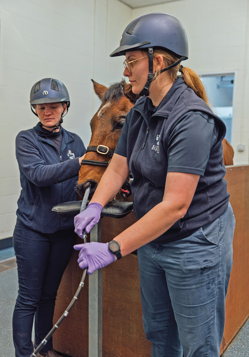
Although videoendoscopes suitable for gastroscopy are commonplace in the UK and Ireland, confidence in, and indeed experience of, the procedure might vary among clinicians. When embarking on gastroscopy in practice, a few simple tips may reduce any anxiety and optimise the diagnostic usefulness of the procedure.
[1] Take a friend
When performing gastroscopy at the yard, try to scope as a pair of vets or with a technician or veterinary nurse. You will be far more effective navigating the stomach with a knowledgeable person passing the scope, and the entire visit will be more efficient. Also, except for professional yards, it asks a lot of our clients to pass a gastroscope into their own horse.
[2] Write a pre-visit checklist
Avoid leaving any essential equipment at home (or at the yard) by writing a checklist to run through before you leave for, and return from, your visit.
[3] Handle with care
Take care of the gastroscope once it is out of its travel case; set everything else up first. Always check where the tip of the gastroscope is, and don’t let it bash against anything or slide off the table/trolley.
[4] Sedate as the final step before scoping
Make sure you have everything ready, switched on, and a working live image from the gastroscope before you sedate the horse. No one wants their horse sedated only to find that an essential connecting wire has been left at the practice, or is malfunctioning, and the procedure has to be aborted.
[5] Protect the scope
Use a naso-oesophageal tube (shortened stomach tube) as a conduit for the gastroscope if your scope is narrow enough, and the patient’s nasal passages are wide enough for the tube. It is impossible to overstate the benefits of using a naso-oesophageal tube in this way (Panel 2). Where this is not achievable, the scope can be protected from chewing by holding the mouth open with a Hausmann’s gag.
Panel 2: Why use a naso-oesophageal tube as a conduit for the gastroscope?
- The scope is protected from retroflexion and cheek tooth chomping, avoiding the use of a dental gag, which is unsightly and a potential weapon.
- The scope passes more freely inside the naso-oesophageal tube, making travel around the stomach much easier.
- Relative to no tube, horses are quiet and comfortable during the procedure because they can’t feel the scope moving in and out of the sensitive nasopharyngeal area.
- No matter what, the horse cannot chew the scope, which reduces everyone’s anxiety.
- For all of the above reasons, the procedure is usually faster.
[6] Hat hair don’t care – keep your team safe
Handlers standing on either side of the horse while assisting in gastroscopy (holding the horse’s head or passing the naso-oesophageal tube if used) are at increased risk of injury, which can be reduced with protective headwear.
[7] Inflation
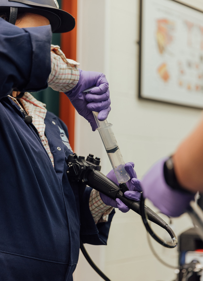
Inflate the stomach just enough to visualise the entrance to the pylorus – it can be harder to navigate down to the pyloric region if the stomach has been widely distended. Be mindful that your finger isn’t stuck down on the “inflate” button due to any nervousness. Most commonly in practice, clinicians will use suction to remove any surplus air left in the stomach, which might reduce the risk of a post-gastroscopy colic (although the evidence of the usefulness of this is lacking).
[8] Delayed gastric emptying
Despite adequate fasting (usually 14 to 16 hours), it can be disappointing to find, in some horses, that their stomach still appears to be full when gastroscopy commences. This may be because feed was given mistakenly, or the horse has consumed bedding or faeces. Both can be avoided with the use of a soft “grazing” muzzle during the overnight fast. Sometimes a legitimate delay can be seen in gastric emptying, and further fasting for 4-6 hours may be enough to complete the procedure. Should this not be the case, the horse might have a gastric impaction; further discussion on gastric impactions can be found elsewhere3.
[9] Duodenal examination
Where there is a clinical concern, such as weight loss, hypoalbuminaemia, colic, gastric impaction or delayed gastric emptying, it can be helpful to assess the mucosal appearance of the duodenum. Sometimes, mucosal pinch biopsies are collected, although the diagnostic usefulness of such small samples is limited. Most clinicians would not consider “duodenoscopy” an essential step for every gastroscopy without seeing a prior clinical indication.
[10] Oesophageal examination
Do not forget to check the mucosal appearance of the distal oesophagus as the scope is removed from the stomach. Ulceration can be found at this site, although probably not as commonly as gastric locations. It is often easier to assess the oesophagus with gentle inflation alongside withdrawal of the scope, compared to the unfurling of the oesophagus “on the way in”.
To learn more about EGUS, listen to the new EGUS podcast series from GastroGard®. Veterinary experts in this field cover key topics, including Rose Tallon discussing why glandular ulcers can be so difficult to treat, the author (Victoria South) covering the importance of management and nutrition when treating gastric ulcers, and Michael Hewetson discussing gastric ulcer syndrome in the racehorse. Available on the Boehringer Academy (www.boehringer-academy.co.uk)
- An educational service sponsored by Boehringer Ingelheim, the makers of GastroGard® 370 mg/g oral paste. Further information available in the SPC or from Boehringer Ingelheim Animal Health UK Ltd., RG12 8YS, UK. GastroGard® is a registered trademark of Boehringer Ingelheim Animal Health France SCS, used under licence. ©2024 Boehringer Ingelheim Animal Health UK Ltd. All rights reserved. Date of preparation: January 2024. UI-EQU-0006-2024. Use Medicines Responsibly.
References
- Sykes BW, Hewetson M, Hepburn RJ, Luthersson N and Tamzali Y (2015). European College of Equine Internal Medicine Consensus Statement – equine gastric ulcer syndrome in adult horses, J Vet Intern Med 29(5): 1,288-1,299.
- The Equine Gastric Ulcer Council (1999). Recommendations for the diagnosis and treat- ment of equine gastric ulcer syndrome (EGUS): The Equine Gastric Ulcer Council, Equine Vet Educ 11(5): 262-272.
- Talbot SE, Tallon R and Dunkel B (2023). Clinical presentation and outcome of gastric impactions with or without concurrent intestinal lesions in horses, J Vet Intern Med 37(4): 1,544-1,551.